Month: July 2022

Greater trochanteric pain syndrome – Wilson Tang

Greater trochanteric pain syndrome

Do you experience pain over the side of your hip/pelvis? Do your symptoms worsen when sleeping on your sides, or after performing physical activity such as jogging or walking up and down stairs? It could be Greater Trochanteric Pain Syndrome (GTPS).

What is GTPS?

GTPS is a relatively common condition where one feels pain over the side of their hip. These symptoms can often travel down the side of the leg or around the buttock region. It is thought that GTPS occurs due to altered biomechanics and reduced load capacity of the hip muscles. These muscles provide stability during standing, walking and other movements. GTPS may feel worse when first waking in the morning, during movement, or even after being sedentary for some time.


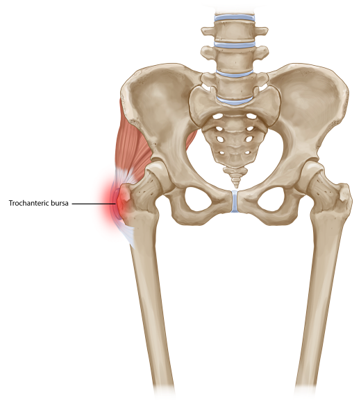
The components of GTPS can often be separated into 1) gluteal tendinopathy (often termed tendinosis if confirmed with ultrasound imaging), and 2) with or without greater trochanteric bursitis (again can be seen via ultrasound imaging). Gluteal tendinopathy simply refers to the musculotendinous component of the condition, and bursitis simply refers to the inflamed area of cushioning over the bony part of the hip area (see Figure 1.)


Who’s at risk?

GTPS is often seen in women above the age of 35-40 years, but can occur earlier and to anyone. Individuals often report a gradual progression of symptoms, which may be preceded by a specific change in activity or exact moment of injury. For example, you may have started running again, or you slipped and bumped your low back/hips, or you may have even been out with the flu and have been laying on the couch a bit more than usual.


Can we diagnose GTPS without imaging?

Diagnosing GTPS can be done quite accurately by a physiotherapist when using a combination or cluster of tests to improve sensitivity and specificity. For example, palpation of the greater trochanter and any combination of loading/compressive testing of the lateral hip structures/muscles (30 sec SLS test, FABER, FADER-R etc) gives the best diagnostic accuracy. It is important to distinguish those with GTPS and those experiencing lumbar spine referred pain, as symptoms can be felt around the same areas but will be managed differently.


Managing GTPS

Whilst it may be tempting to push through the pain, appropriate management may require certain activity and postural modifications to keep pain minimal.

This may include:

Sleep habits

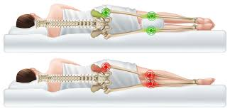
  • Sleeping on your back or front if possible. Sleeping on sides increases compression over the sore irritated bursa/muscle tendons and can further aggravate symptoms.

  • If sleeping on your sides, a softer mattress (mattress toppers are a great idea) may help reduce the compressive pressure over the sore area, or sleeping with a pillow between the knees may also help reduce compression over the sore area.


Sitting/standing postures

  • Avoid sitting with legs crossed where possible, as this further increases pressure over the sore area.

  • A higher chair height typically helps reduce the angle between your legs and pelvis, which may help reduce pressure over the side of your hips (try sitting on top of a pillow at the kitchen table).

  • Avoid putting more weight on the sore leg when standing still a.k.a. “Hip hanging”.


Walking/stairs

  • Walking uphill and stairs often increases loading and compression over the lateral hip muscles. If possible ease up on these activities for a short period to allow symptoms settle.


Medications

  • Oral anti-inflammatories may help the process along, and allow for you to get started on the active exercise and rehabilitation.

  • Local steroid injections may help relieve symptoms short-term, but there is some emerging research questioning the long-term effectiveness and safety of receiving too many steroid injections


Manual therapy

  • Some treatment directed over muscles involved may help relieve symptoms short term, but typically we avoid direct pressure over the bony aspect of the outer hip.

  • Taping may also be an option to help offload sore structures during movement.


EXERCISE

As with many injuries/conditions, exercise is key for long-term management of GTPS. Specifically targeting the muscles of the hip girdle, and addressing biomechanical factors will help resolve symptoms in the long run. However, knowing how much to do and what to do specifically is crucial as always. For example, walking 10 mins compared to 30 mins can be a completely different ball park for the affected muscles/structures, and may be the difference between a manageable exercise amount and an amount that makes symptoms worse.


A gradual loading program to strengthen those muscles of the pelvic girdle, and offloading sore structures with appropriate rest or modifications will get you feeling much better and get you back to doing what you love.

Assessment, Management and Prevention of Calf Muscle Strain Injuries – Review Matt Delaney

Research Review:


The Assessment, Management and Prevention of Calf Muscle Strain Injuries: A Qualitative Study of the Practices and Perspectives of 20 Expert Sports Clinicians


https://www.ncbi.nlm.nih.gov/pmc/articles/PMC8761182/


This paper aimed to evaluate the current practices regarding assessment, management and prevention of calf muscle injuries. This was done through interviews with 20 expert clinicians who either work in elite sport or were researchers in the relevant field.


The first portion of the interviews focused on evaluating injury characteristics and in particular differentiating gastrocnemius strains from soleus strains. There was a consensus that soleus injuries can be an accumulation of symptoms that tend to present as a gradual onset, whether gastrocnemius injuries have a distinct mechanism. Soleus injuries are more common in steady state running whether gastrocnemius injuries tend to involve acceleration, jumping, landing or sprinting. Low grade soleus injuries can be more difficult to localise the source of pain, with focal pain being more common in injuries involving gastrocnemius (or severe soleus injuries).


Risk factors including past history of calf strain or previous lower limb soft tissue injury as well as recent interruptions to or sudden increases in running workloads, this is particularly relevant in soleus injuries. This was noted more often in the pre-season period as athletes were re-introduced to running workloads and higher intensity. In objective testing changing knee position can help differentiate muscle involvement for stretch and strength testing (flexed for soleus and extended for gastrocnemius) however in severe injuries symptoms will present similarly regardless of knee position. Appropriate loading during your examination from low load (isometrics) through to concentric (double and single leg) and plyometric if able assisted in grading the injury severity.


After assessment for injury severity and pathology, experts then looked at rehabilitation. Initial goals were normalising gait pattern and appropriate early loading. Once able to demonstrate single leg calf raise capacity athletes should then be progressed to loaded strengthening, commonly this involved smith machine calf raises, and seated calf raises. These should be set up with parameter reflecting the sport (eg: strength endurance in runners or force generation capacity in rugby or sprinters) and progress through range of motion as the athlete tolerates initially starting on flat then moving to an incline.  Experts also advised to strongly considered soleus capacity in all calf injuries and athletes regardless of the pathology involved prior to introducing dynamic exercise.


The next step is to introduce plyometric/ballistic exercises. Two main exercise streams were identified: (1) repeated stretch-shortening cycle’s (SSC) over small length-excursions associated with a rhythmic muscle tendon unit (MTU) action (e.g. skipping or single leg pogos), and (2) single or several SSCs over larger length excursions (e.g. single leg countermovement jump, forward hopping) associated with an accelerative MTU action. These should be prescribed first in the vertical plane and then in the horizontal plane due to increased tissue demands required in the latter. Plyometric exercise selection should reflect the sporting demands and with sports such as AFL or soccer there may be a need to develop both rhythmic and accelerative plyometrics.


Determining readiness to run was another topic discussed throughout the paper with experts reporting that gait re-training drills were initiated as soon as practical with examples such as stair ascents, bear crawls or wall A-drills used. To be cleared for return to running 3 checkpoints were required, firstly achieving appropriate tissue strength capacity (eg: single leg calf raise endurance or loaded calf strength), secondly the ability to tolerate repeat hopping and finally the absence of any clinical signs or symptoms (pain on stretch, reduced ROM etc). Six recommendations were identified from information provided by experts to guide running rehabilitation: (1) initially run on alternate days, (2) avoid “plodding” early, (3) do not progress volume and intensity on consecutive running days, (4) schedule off-field exercises (e.g. loaded strengthening) after running, (5) shape running progressions to meet the demands of the sport—don’t overshoot with excessive volume, (6) avoid sudden changes in conditions, such as the surface and footwear. The reasoning to avoid plodding during early running rehabilitation was that it had been found to predispose to recurrence for injury involving soleus. Endurance capacity does need to be ticked off however the recommendation was to check this off last once higher speeds and change of direction loads had been hit.


A return to play checklist is detailed below, in terms of strength tests Smith machine raises (extended knee) at least 1 x bodyweight for 5 repetitions and seated calf raises 1.5 x bodyweight for 5 repetitions were recommended. For vertical hop tests a countermovement or depth jump was used and for horizontal capacity a single hop and triple hop test were used with an asymmetry of <10% compared to the unaffected side. Once the athlete has returned to sport, these tests can also be used as a monitoring criteria in regards to injury prevention. It was found the risk of recurrence was due to four main factors: (1) Increased chronological age, (2) previous calf muscle injury, (3) previous lower limb injury and (4) exposure/loading history. Experts agreed that the best way to mitigate risk was to ensure uninterrupted sports exposure whilst monitoring player training loads and continuing to hit strength benchmarks.

The necessary evil of pain – Monique Rowlands

Let’s talk about the necessary evil of acute pain.

Acute pain is something that we have all experienced at one point or another. Often, patients find the pain of their injury quite distressing, which is fair. Pain is a noxious stimulus, it excites the alarm centres in the brain, setting off a series of protective mechanisms including a psychological distress response. However, I want to spend some time thinking about how acute pain can be used as a training tool to fix bad habits, learn new things about exercise or training loads and be beneficial in our experience of life.

First, let’s consider what I mean by bad habits. Often acute pain is due to tissue stress, caused by abnormal loading of muscles or joints – bad habits or poor biomechanics. Our bodies are amazing things and are extremely patient with our bad habits and poor techniques. Bodies have a biomechanical tolerance, meaning that you can perform an activity with bad technique without experiencing pain up to a certain point. This point is different for every body, some may reach it sooner than others, but at some point, tissue loading reaches a point where the joint/ ligament/muscle is no longer able to tolerate the abnormal biomechanical loading. This point is where tissue becomes pain sensitized by an inflammatory response and requires treatment/adaptation to the mechanics to settle down.

To explain this using a real-life example, let’s talk about knee pain. A patient may present with a 3-month history of knee pain that is worse with running, squats, lunges, and stairs. They have recently stopped exercising because of the pain and are feeling quite down about the impact the pain has in their life. Prior to the onset of pain, they ran 5km a day, even though they ran with a “knock-knee” pattern. A month before the pain started, the patient started a gym class with lots of squats and lunges. In this example, the patient is experiencing a condition called Patello-Femoral Joint (PFJ) pain, which is typically caused by poor biomechanics of the knees. In this case, the patient’s knees tolerated 5km of running with poor biomechanics for years before any symptoms arose. It was only with the increased load of squats and lunges that the knees became painful during their regular activities like running and stairs.

The patient can utilise their pain to their benefit, by using it to correct the longstanding biomechanical flaw in their running and squatting pattern. When the knee is positioned properly, the patient will be able to significantly reduce their pain. This is a helpful way to use pain. It becomes a feedback tool for the patient in helping them learn new movement patterns that will ultimately reduce the recurrence of pain in the future. Better movement patterns lead to better performance, stronger bodies and less wear on joints. Therefore, pain is a helpful tool in life. It is unavoidable, everybody feels acute pain and with the correct management and guidance from your local physio can assist in making you better at what you do.