Author: Michael Ranger

Bone Stress Injuries in Runners and Athletes – Isaac McMillan

Understanding Bone Stress Injuries

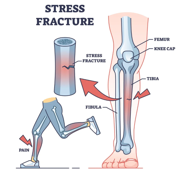
Bone Stress Injuries (BSIs) are common overuse injuries that occur when a bone is unable to withstand repetitive mechanical loading. They develop when microscopic damage accumulates faster than the body can repair it through normal bone remodeling.

Rather than occurring suddenly like a traumatic fracture, BSIs exist on a continuum. The process typically begins as a stress reaction, progresses to a stress fracture, and in severe cases can develop into a complete fracture.

BSIs are particularly common in runners, track and field athletes, military recruits, and athletes involved in high-volume training. Research suggests that approximately one-third of long-distance runners will experience a bone stress injury at some point during their sporting career.

—————————————————————————————–

Why Do Bone Stress Injuries Occur?

Bone is constantly adapting to the loads placed upon it. When training loads exceed the bone’s capacity to recover and remodel, microdamage accumulates.

Several factors influence the risk of developing a BSI:

Training and Biomechanical Factors

Rapid increases in training volume, intensity, or frequency are common contributors. Changes in running surfaces, footwear, or training programs can also alter the forces experienced by the skeleton.

Muscles play an important role in absorbing shock during running. Muscle weakness or fatigue reduces this shock-absorbing capacity, increasing the load transferred to the bone.

Bone Capacity

An athlete’s history of physical activity influences bone strength. Individuals with a long history of weight-bearing exercise generally have stronger bones and are less susceptible to injury.

Nutrition is equally important. Low energy availability, inadequate calorie intake, and conditions such as Relative Energy Deficiency in Sport (RED-S) can impair the body’s ability to maintain and repair bone tissue. Female athletes are particularly vulnerable due to the effects of low energy availability on menstrual function and bone health.

Adequate calcium and vitamin D intake is also essential. Calcium contributes to bone rigidity, while vitamin D helps the body absorb calcium efficiently.

—————————————————————————————–

Common Symptoms

Bone stress injuries typically develop gradually and are often linked to a change in training during the preceding six to eight weeks.

Athletes commonly report:

• A dull ache during running or sport

• Symptoms that resolve when exercise stops

• Increasing pain with continued training

• More localised tenderness as the injury progresses

As the condition worsens, pain may occur earlier during exercise, persist afterwards, or even be present during walking. Resting pain or night pain may indicate a more serious injury and should be assessed promptly.

A key clinical finding is localised bony tenderness, often within a small area less than 5 cm in diameter.

—————————————————————————————–

Diagnosis

Early diagnosis is critical and is associated with a faster and more successful recovery.

Physiotherapists use a combination of clinical history, palpation, and bone loading tests such as hopping or functional impact assessments. If a bone stress injury is suspected, MRI is considered the gold standard imaging modality due to its high sensitivity and ability to identify injuries before a fracture develops.

—————————————————————————————–

Treatment

The primary goal of treatment is to allow the injured bone to heal while maintaining overall fitness and strength.

Most low-risk bone stress injuries require a temporary reduction or cessation of running. Pain is the key guide throughout rehabilitation. Walking and daily activities should be pain-free before running resumes.

Strength training can often begin early and may include:

• Calf strengthening • Hip and knee strengthening

• Core stability exercises

• Foot intrinsic strengthening

Maintaining cardiovascular fitness is also important. Activities such as swimming, cycling, and deep-water running can help preserve aerobic capacity while reducing bone loading.

—————————————————————————————–

Returning to Running

A gradual return-to-running program should only begin once walking is pain-free for at least five consecutive days.

Initially, athletes should run every second day and start at approximately 50% of their normal pace and distance. Running volume should increase before intensity.

Importantly, bone recovery occurs more slowly than cardiovascular fitness gains. Athletes often feel fit enough to progress faster than their bones can tolerate. For this reason, progression should always be guided by symptoms rather than fitness levels.

If symptoms return during rehabilitation, training should be reduced and reassessed before progressing further.

—————————————————————————————–

Preventing Future Bone Stress Injuries

Successful long-term management requires identifying and addressing the factors that contributed to the injury. This may include optimising training loads, improving strength, addressing nutritional deficiencies, and reviewing running mechanics where appropriate.

While gait retraining may be beneficial in athletes with recurrent bone stress injuries, unnecessary changes to running technique can overload other tissues and potentially create new problems. Any modifications should be carefully prescribed and monitored by a qualified clinician.

With early diagnosis, appropriate load management, and a structured rehabilitation program, most athletes can successfully return to running and reduce their risk of future bone stress injuries.

—————————————————————————————–

References

Warden, S. J., Davis, I. S., & Fredericson, M. (2014).  Management and Prevention of Bone Stress Injuries in Long-Distance Runners. Journal of Orthopaedic & Sports Physical Therapy, 44(10), 749–765. https://doi.org/10.2519/jospt.2014.5334

Warden, S. J., Edwards, W. B., & Willy, R. W. (2021). Optimal Load for Managing Low-Risk Tibial and Metatarsal Bone Stress Injuries in Runners: The Science Behind the Clinical Reasoning. Journal of Orthopaedic & Sports Physical Therapy, 51(7), 1–28. https://pubmed.ncbi.nlm.nih.gov/33962529/

Warden, S. J., Edwards, W. B., & Willy, R. W. (2021). Preventing Bone Stress Injuries in Runners with Optimal Workload. Current Osteoporosis Reports, 19(3). https://pubmed.ncbi.nlm.nih.gov/33635519/
Dizziness and Vertigo – Sophie Knight

Physiotherapists see all kinds of musculoskeletal conditions in the clinic, but not many people are aware that we can also treat some kinds of dizziness and vertigo.

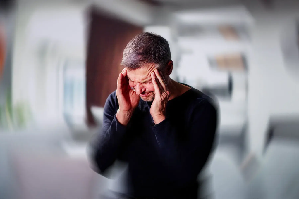
Definition of Vertigo: the sensation that the environment around you is spinning. Vertigo can be associated with nausea, eye flickering, balance difficulties and ringing in the ears.

Vertigo is a symptom that can be caused by several different conditions, and different conditions require different management options. Two common causes of vertigo that physiotherapists can manage includes your inner ear and your neck.

  1. Benign Paroxysmal Positional Vertigo (BPPV)

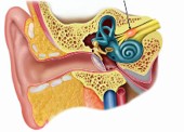
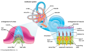
The inner ear is a complex structure made of a maze of canals. Inside the canals are little crystals that are embedded in a bed of jelly. When your head turns, the crystals wobble the jelly which send signals to tell the brain that you are moving.

Sometimes these crystals become dislodged and start rolling around. This will manifest as feeling vertigo for a few seconds when you lay down in bed or roll over. This type of vertigo is commonly associated with flickering eyes when the vertigo happens.

BPPV is managed with an easy technique called the Epley’s manoeuvre. This is completed at least once to help move the ear crystals back into the right spot (Hilton et al., 2014). The patient can be taught how to complete this technique at home to ensure that if they have subsequent instances of BPPV, they are equipped with the tools to self-manage this condition.

In cases where Epley’s manoeuvre is contraindicated or not fully successful, vestibular rehabilitation can be completed, where tailored exercises are prescribed to re-train the vestibular system to get used to sudden movements again (Bressi et al., 2017).

  1. Vestibular Neuritis

This is a condition where the nerve that connects to your ear becomes inflamed and results in vertigo, the sensation of full ears, balance changes and nausea. It is seen to come on before or after a viral infection. Compared to BPPV where symptoms occur with movement and resolve quickly, vestibular neuritis vertigo tends to be more constant. A GP can prescribe different medications to help with reducing nausea and inflammation. Concurrently a physiotherapist will prescribe vestibular rehabilitation to gradually expose the vestibular system to the things that are most aggravating (Tokle et al., 2020).


  1. Cervical spine related dizziness

Another cause of vertigo is related to your cervical spine, or your neck. Due to the complex nature of the nerves that exit your neck, if there is neck stiffness, pain, or other impairments, it can result in changing the way the signals are sent to your brain.  It can also be associated with headaches. This form of dizziness will feel different and less “violent” compared to BPPV or vestibular neuritis, and it can last a few minutes up to a few hours at a time (Wrisley ey al., 2000). Aggravating factors might include prolonged head postures.

Cervicogenic dizziness can be managed in different ways. Physiotherapists will often use manual or “hands-on” therapy to help with symptom management. From there, exercises will be used to address deficits in the strength, endurance or control of the neck, in addition to exposure to aggravating factors once the patient is ready (De Vestel et al., 2022).


References

Bressi, F., Vella, P., Casale, M., Moffa, A., Sabatino, L., Lopez, M. A., … & Sterzi, S. (2017). Vestibular rehabilitation in benign paroxysmal positional vertigo: Reality or fiction?. International journal of immunopathology and pharmacology30(2), 113-122.

De Vestel, C., Vereeck, L., Reid, S. A., Van Rompaey, V., Lemmens, J., & De Hertogh, W. (2022). Systematic review and meta-analysis of the therapeutic management of patients with cervicogenic dizziness. Journal of Manual & Manipulative Therapy30(5), 273-283.

Hilton, M. P., & Pinder, D. K. (2014). The Epley (canalith repositioning) manoeuvre for benign paroxysmal positional vertigo. Cochrane database of systematic reviews, (12).

Tokle, G., Mørkved, S., Bråthen, G., Goplen, F. K., Salvesen, Ø., Arnesen, H., … & Wilhelmsen, K. T. (2020). Efficacy of vestibular rehabilitation following acute vestibular neuritis: a randomized controlled trial. Otology & Neurotology41(1), 78-85.

Wrisley, D. M., Sparto, P. J., Whitney, S. L., & Furman, J. M. (2000). Cervicogenic dizziness: a review of diagnosis and treatment. Journal of Orthopaedic & Sports Physical Therapy30(12), 755-766.

Living Well with Knee Osteoarthritis

Understanding, Managing, and Moving Forward

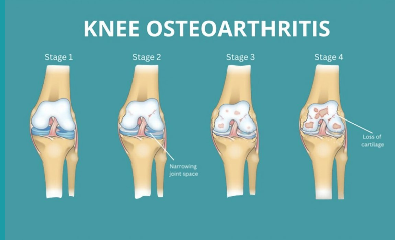
Knee osteoarthritis (OA) is a common condition that can cause ongoing pain, stiffness, and difficulty with everyday activities. It affects the whole joint — including cartilage, bone, ligaments, and muscles — and is part of your body’s active response to small joint injuries over time. While it can be uncomfortable, OA is manageable and doesn’t always get worse. With the right approach, most people continue to live active, fulfilling lives.

Diagnosis and Outlook

Doctors often diagnose knee OA based on symptoms such as pain lasting more than three months, morning stiffness under 30 minutes, and difficulty with movement. X-rays are usually unnecessary because they don’t reliably reflect pain levels or guide treatment. For most people, OA remains stable, with occasional flare-ups that settle with time. Only a small number of people will ever need a knee replacement.


The Importance of Physical Activity

Many people with knee OA reduce their activity levels out of fear of making the pain worse. In fact, avoiding movement often increases stiffness, weakness, and discomfort over time. Staying active is one of the best things you can do for your joints, muscles, and overall health.

You don’t need intense workouts — small, regular movements make a big difference. Moderate-intensity activity means you’re breathing a little harder but can still speak in sentences. Examples include brisk walking, swimming, cycling, water aerobics, yoga, or doing household tasks more energetically.

Tips for increasing your daily activity:

  • Take small steps — stairs instead of elevators, park further away, walk while talking on the phone.

  • Avoid long periods of sitting; move every 30–60 minutes.

  • Break activity into short bouts of 10 minutes or more.

  • Gradually increase duration or intensity.

  • Mix activities — walking, gardening, dancing, playing with grandchildren.

  • Track steps or activity using a phone or monitor.

  • Pace yourself and be kind to your body, even on “bad” days.


Managing Your Weight

Being overweight places extra stress on your knees — each kilogram of body weight can add about four kilograms of load on your knees during daily activities. Extra weight can also increase inflammation in the body, making pain and stiffness worse. Losing even a small amount of weight — 5–10% of your body weight — can reduce joint load, improve pain, increase function, and may delay or reduce the need for medications or surgery.

Tips for weight management:

  • Combine regular physical activity with healthy eating habits.

  • Set realistic goals and be patient; don’t give up if progress is slow.

  • Eat mindfully: slow down, savour meals, and avoid eating when bored or stressed.

  • Plan meals and snacks ahead, and keep a food diary.

  • Drink water regularly and aim for sufficient sleep.

  • Learn about healthy food choices and portion sizes.

  • Engage friends, family, or a buddy for support and accountability.

  • Handle holidays or special events by planning ahead — enjoy the event but make mindful choices.

Key points:

  • Weight loss can noticeably reduce knee pain and improve function.

  • Aim for 5–10% of your body weight to see meaningful benefits.

  • Combining diet and exercise gives the best results.


Overcoming Common Barriers

Everyone faces challenges staying active or managing weight. Creative solutions can help:

  • Time: Schedule activity like an appointment; short bursts count.

  • Pain: Mild discomfort is normal; severe or prolonged pain requires adjustment.

  • Fatigue: Exercise often increases energy and improves sleep.

  • Motivation: Track progress, reward yourself, exercise with friends.

  • Stress or emotional eating: Try music, social support, mindfulness, or walking instead of using food as a coping strategy.


The Bottom Line

Knee osteoarthritis does not mean you have to stop doing the things you love. Staying active, managing your weight, learning to manage pain, and seeking support from health professionals can help you live well with OA. Every small step counts toward stronger knees, reduced pain, and better overall health.

For additional information:

https://www.myjointpain.org.au

https://arthritisaustralia.com.au

https://www.msk.org.au/diet/

https://www.healthdirect.gov.au/sleep

Tibialis Posterior Tendinopathy

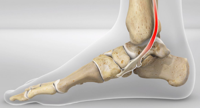
The tibialis posterior (TibP) muscle runs along the inside of your shin and down to your mid foot. It functions to invert and plantarflex the foot and control your arch. This means it plays a big role with walking, running and balancing.

TibP tendinopathy will present with pain and stiffness on the inside of your ankle and/or foot. There may also be swelling present. TibP tendinopathy pain will warm up with activity but often feels worse after a period of resting. It is typically associated with an increase of activity or a recent change in activity.

Examples might include:
– Someone who just took up running or increased their km per week
– An athlete bought a new pair of shoes for their sport
– A switch to synthetic turf when historically playing on grass

Tendons stem from our muscles which attach them to bone. Tendinopathy is considered an overuse injury of tendons. This means that the tendon is not coping with the demands that is being placed on it. This leads to pain and dysfunction of that tendon.

Assessment
Physiotherapists will ask about the history of the pain which will provide clues into whether it is or isn’t TibP tendinopathy.

A physical exam will also be performed which may include:
– Double or single leg calf raise
– Jumping or hopping
– Muscle strength testing
– Palpation of the lower leg

Imaging?

Imaging (ultrasound or MRI) is not typically required for accurate diagnosis of this injury. This is because imaging is poorly correlated with symptoms; a study found that under ultrasound imaging, 48% of people showed TibP tendinopathy despite have no medial foot or ankle pain (Mills et al., 2020).

Some people also present with normal imaging despite having clinical
features of TibP tendinopathy. Imaging is likely only required if your physiotherapist suspects a bony injury.

Treatment and Management
TibP tendinopathy can be successfully managed with physiotherapy. It may involve a period of relative rest and concurrent strengthening exercises of the TibP and other muscles in the body.

– Isometric ankle inversion
– Single leg calf raises
– Eversion
– Toe raises
– Energy storage and release exercises (hopping, depth jumps, lateral bounds etc)
– Strengthening of proximal muscles (hip and knee strength)

Other management strategies involve:
– Topical or oral NSAIDs (anti-inflammatory medication)
– Soft tissue release of TibP and other muscles in the lower leg
– Joint mobilisations

Differential Diagnoses – If it isn’t TibP tendinopathy, what else could it be?
– Deltoid ligament injury
– Navicular bone injury
– Flexor hallucis longus tendinopathy
– Tarsal tunnel syndrome

Apophysitis Injuries in Children

The apophysis is a normal outgrowth on long bones where tendons or ligaments attach. These areas will continue to fuse and harden as children get older and their growth plates fuse.

Apophysitis is a condition of overuse and inflammation at these apophysis sites. It is the result of repetitive stress from activities/sport. These injuries will worsen with activity and improve with rest. Since the apophysis growth plate is still open, combined with the repetitive action of muscles, this makes these areas especially vulnerable to injury.

Some common overuse apophysis injuries include:
1. Osgood-Schlatter’s Syndrome (knee)
2. Sever’s Syndrome (heel)
3. Little League Elbow (elbow)
4. Iselin’s Syndrome (outside of the midfoot)

Osgood-Schlatter’s
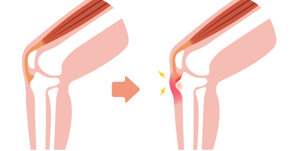
This is apophysitis at the tibial tuberosity, or the bony bump at the top of your shin. This is where the quadriceps muscle attaches onto the shin via the patella tendon.

Signs and Symptoms
– Pain and/or swelling at the tibial tuberosity
– Pain at the knee when participating in sport/activity, especially running and jumping
– A recent growth spurt
– Symptoms resolve with rest

Sever’s
This is apophysitis of the calcaneus (heel bone). This is where the gastrocnemius and soleus muscles attach onto the foot via the Achilles tendon.

Signs and Symptoms

– Pain at the heel when participating in sport/activity, especially with running and jumping

– Pain and/or swelling at the heel

– A recent growth spurt

– Symptoms resolve with rest

Assessment
Physiotherapy assessment will begin with a subjective interview to gain insight into the history of the pain to determine if it is or isn’t apophysitis. Your GP may have requested an X-ray of the area which can help determine diagnosis, but it will sometimes come back normal.

The physiotherapist will look at:
– Squats
– Jumps and hops
– Calf raises
– Palpation of the painful area
– Muscle strength and length testing

Management
Apophysitis generally starts with a period of relative rest. Depending on the aggravating activities, other sports may be tolerated better in the meantime. For example, instead of going to soccer, riding a bike or swimming may be less painful.

Strengthening and stretching exercises are also essential to managing apophysitis. This might include stretching the quadriceps, hamstrings, calves and glutes. Strengthening of the affected muscles will also be key.

– Double leg glute bridges
– Theraband quad extensions
– Hip abduction exercises
– Squats or lunges
– Double or single leg calf raises

Other management strategies may include:
– Soft tissue mobilisation of the affected muscles

– Foam rolling or spikey ball at home

– Movement retraining

– Rigid taping or KT taping of the affected area for symptomatic relief

Hamstring injuries in football – Wilson Tang

Hamstring injuries can often be a footy player’s worst nightmare. Due to the nature of the game, AFL and hamstring injuries go hand in hand. Common ways to injure hamstrings include sprints, kicking, and jumping which often involve a rapid stretch and/or contraction of the muscle. If not picked up early and managed well, they can often be an ongoing battle for athletes throughout the season, and in more serious cases place athletes on the sidelines.
How to identify a hamstring injury?
Significant hamstring injuries may be easy to detect, with characteristic “popping” sensation often described. Associated pain, bruising, swelling, weakness or limited hamstring flexibility may also be tell-tale signs. Naturally, a previous hamstring injury instantly increases your suspicions.
However, mild injuries may be trickier to pick up. Additionally, other structures around the hamstrings may be the culprit such as the adductor/groin muscles, sciatic nerve irritation, low back pain referral etc.
A few simple tests may be used to test the hamstring muscle contraction and range:
  • Shoe off / floor swipe test.
  • Prone knee bend.
  • Single-leg bridge.
  • Arabesque / single-leg deadlift.
  • MHFAKE (maximum hip flexion, active knee extension).
If any tests reproduce familiar pain then further assessment may be warranted.
Rehab tips
Appropriate rest, gradual loading, and sports-specific rehab are all common approaches for hamstring recovery. However, it’s important to consider other areas around the hamstrings during rehab to prevent ongoing recurrence. All structures above, around, and below the hamstrings can influence risk of injury and should be assessed and targeted accordingly.
If you’re suspicious of a hamstring niggle or are struggling with recurrent hamstring injuries, have a chat to your local physiotherapist about how best to approach rehab management.