
Understanding Bone Stress Injuries
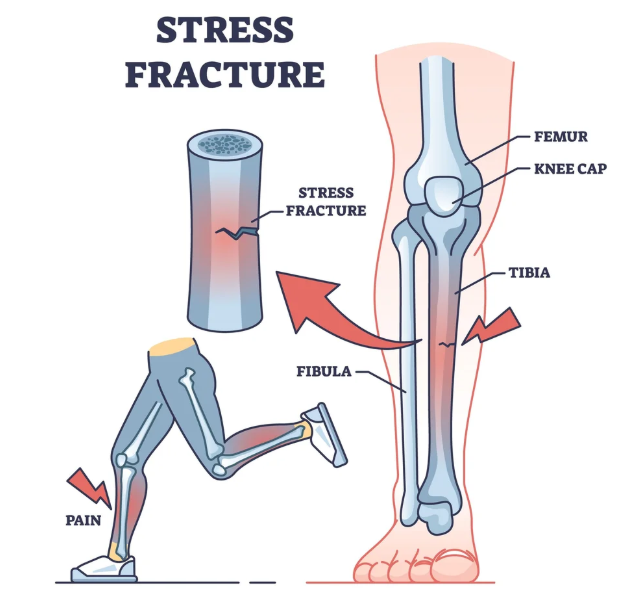
Bone Stress Injuries (BSIs) are common overuse injuries that occur when a bone is unable to withstand repetitive mechanical loading. They develop when microscopic damage accumulates faster than the body can repair it through normal bone remodeling.
Rather than occurring suddenly like a traumatic fracture, BSIs exist on a continuum. The process typically begins as a stress reaction, progresses to a stress fracture, and in severe cases can develop into a complete fracture.
BSIs are particularly common in runners, track and field athletes, military recruits, and athletes involved in high-volume training. Research suggests that approximately one-third of long-distance runners will experience a bone stress injury at some point during their sporting career.
—————————————————————————————–
Why Do Bone Stress Injuries Occur?
Bone is constantly adapting to the loads placed upon it. When training loads exceed the bone’s capacity to recover and remodel, microdamage accumulates.
Several factors influence the risk of developing a BSI:
Training and Biomechanical Factors
Rapid increases in training volume, intensity, or frequency are common contributors. Changes in running surfaces, footwear, or training programs can also alter the forces experienced by the skeleton.
Muscles play an important role in absorbing shock during running. Muscle weakness or fatigue reduces this shock-absorbing capacity, increasing the load transferred to the bone.
Bone Capacity
An athlete’s history of physical activity influences bone strength. Individuals with a long history of weight-bearing exercise generally have stronger bones and are less susceptible to injury.
Nutrition is equally important. Low energy availability, inadequate calorie intake, and conditions such as Relative Energy Deficiency in Sport (RED-S) can impair the body’s ability to maintain and repair bone tissue. Female athletes are particularly vulnerable due to the effects of low energy availability on menstrual function and bone health.
Adequate calcium and vitamin D intake is also essential. Calcium contributes to bone rigidity, while vitamin D helps the body absorb calcium efficiently.
—————————————————————————————–
Common Symptoms
Bone stress injuries typically develop gradually and are often linked to a change in training during the preceding six to eight weeks.
Athletes commonly report:
• A dull ache during running or sport
• Symptoms that resolve when exercise stops
• Increasing pain with continued training
• More localised tenderness as the injury progresses
As the condition worsens, pain may occur earlier during exercise, persist afterwards, or even be present during walking. Resting pain or night pain may indicate a more serious injury and should be assessed promptly.
A key clinical finding is localised bony tenderness, often within a small area less than 5 cm in diameter.
—————————————————————————————–
Diagnosis
Early diagnosis is critical and is associated with a faster and more successful recovery.
Physiotherapists use a combination of clinical history, palpation, and bone loading tests such as hopping or functional impact assessments. If a bone stress injury is suspected, MRI is considered the gold standard imaging modality due to its high sensitivity and ability to identify injuries before a fracture develops.
—————————————————————————————–
Treatment
The primary goal of treatment is to allow the injured bone to heal while maintaining overall fitness and strength.
Most low-risk bone stress injuries require a temporary reduction or cessation of running. Pain is the key guide throughout rehabilitation. Walking and daily activities should be pain-free before running resumes.
Strength training can often begin early and may include:
• Calf strengthening • Hip and knee strengthening
• Core stability exercises
• Foot intrinsic strengthening
Maintaining cardiovascular fitness is also important. Activities such as swimming, cycling, and deep-water running can help preserve aerobic capacity while reducing bone loading.
—————————————————————————————–
Returning to Running
A gradual return-to-running program should only begin once walking is pain-free for at least five consecutive days.
Initially, athletes should run every second day and start at approximately 50% of their normal pace and distance. Running volume should increase before intensity.
Importantly, bone recovery occurs more slowly than cardiovascular fitness gains. Athletes often feel fit enough to progress faster than their bones can tolerate. For this reason, progression should always be guided by symptoms rather than fitness levels.
If symptoms return during rehabilitation, training should be reduced and reassessed before progressing further.
—————————————————————————————–
Preventing Future Bone Stress Injuries
Successful long-term management requires identifying and addressing the factors that contributed to the injury. This may include optimising training loads, improving strength, addressing nutritional deficiencies, and reviewing running mechanics where appropriate.
While gait retraining may be beneficial in athletes with recurrent bone stress injuries, unnecessary changes to running technique can overload other tissues and potentially create new problems. Any modifications should be carefully prescribed and monitored by a qualified clinician.
With early diagnosis, appropriate load management, and a structured rehabilitation program, most athletes can successfully return to running and reduce their risk of future bone stress injuries.
—————————————————————————————–
References
Warden, S. J., Davis, I. S., & Fredericson, M. (2014). Management and Prevention of Bone Stress Injuries in Long-Distance Runners. Journal of Orthopaedic & Sports Physical Therapy, 44(10), 749–765. https://doi.org/10.2519/jospt.2014.5334
Warden, S. J., Edwards, W. B., & Willy, R. W. (2021). Optimal Load for Managing Low-Risk Tibial and Metatarsal Bone Stress Injuries in Runners: The Science Behind the Clinical Reasoning. Journal of Orthopaedic & Sports Physical Therapy, 51(7), 1–28. https://pubmed.ncbi.nlm.nih.gov/33962529/
Warden, S. J., Edwards, W. B., & Willy, R. W. (2021). Preventing Bone Stress Injuries in Runners with Optimal Workload. Current Osteoporosis Reports, 19(3). https://pubmed.ncbi.nlm.nih.gov/33635519/






Recent Comments