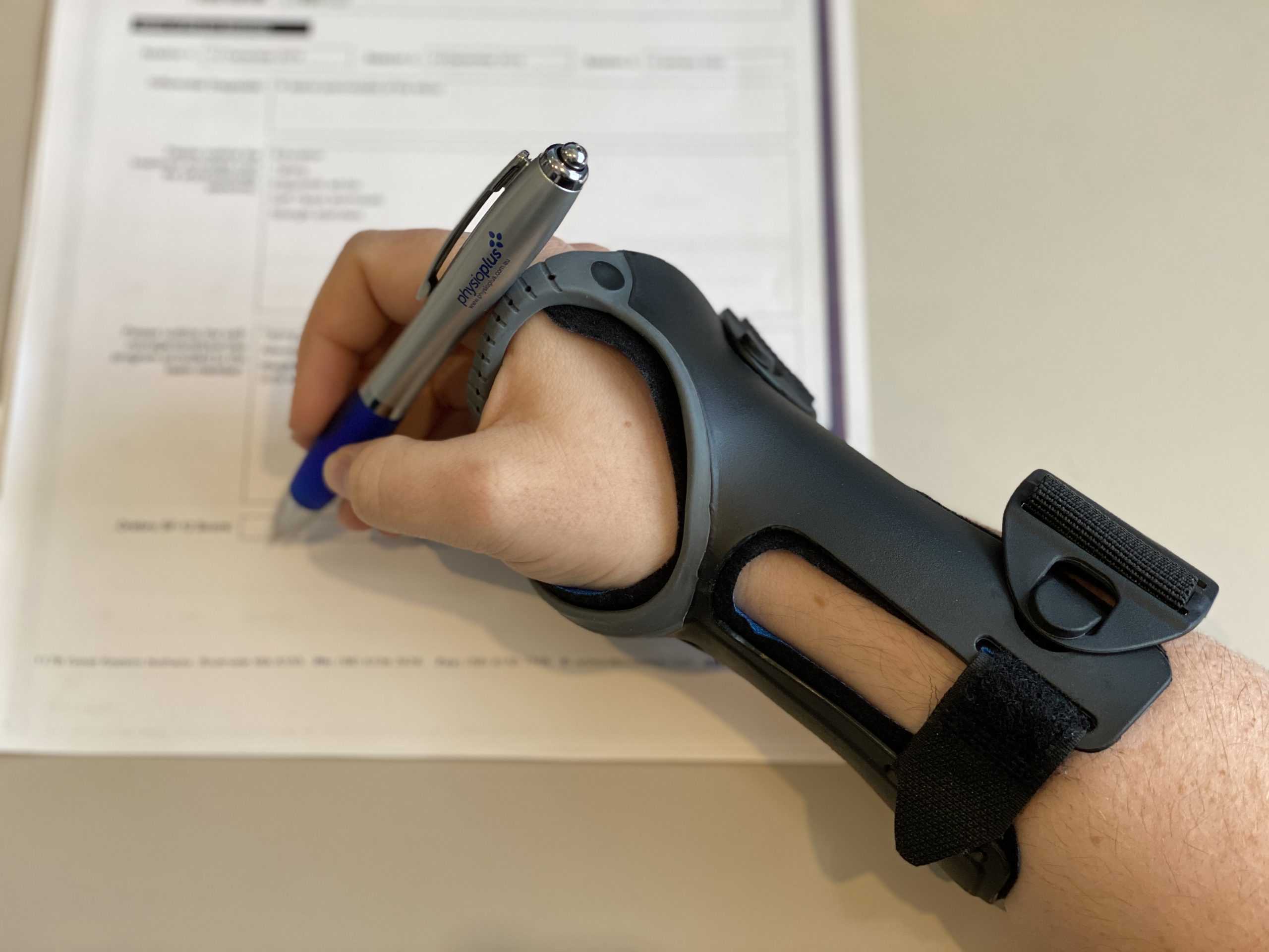
Author: Michael Ranger

Sinus Tarsi – Eliza Osborn

Sinus Tarsi

The sinus tarsi is a tunnel that lies between the talus and calcaneus (heel bone) and
contains structures that contribute to the stability and proprioception of the ankle.
Together, these bones form the subtalar joint which is usually a very stable joint with
minimal movement during weight bearing tasks.

Sinus tarsi syndrome is most often caused by a traumatic ankle injury such as an
inversion ankle sprain or overuse from repetitive activity such as standing or walking.
These injuries can cause instability of the subtalar joint and this excessive movement of
the joint increases the forces placed through the sinus tarsi, triggering an inflammatory
response. It can also occur due to an over-pronated (flat) foot which can cause
compression in the sinus tarsi. Inflammation of the sinus tarsi will usually produce
localized pain over the anterolateral aspect of the ankle (over the site of the sinus tarsi)
and will often be exacerbated by standing or walking/running particularly on uneven
ground.

Athletes with sinus tarsi syndrome will often initially respond well to anti-inflammatory
medication and taping to increase the stability of the subtalar joint which will allow them
to manage their symptoms without significant disruption to their training load. A
graduated strengthening program particularly for the peroneal and calf muscles is
important alongside proprioceptive and balance exercises to reduce the chances of
re-injury.

If you’ve suffered an ankle injury and are experiencing pain, go and visit your
physiotherapist today and they can help you get back on track and fit and firing!

https://www.ncbi.nlm.nih.gov/pmc/articles/PMC2953318/

Ischiofemoral Impingement – Wilson Tang

Ischiofemoral impingement

What is it:
– Narrowing of the space between the lesser trochanter of the femur and ischium of the pelvis

Presentation:
– Pain often reported in the lower buttock region, groin/inner thigh +/- snapping/clunking sensation
– Close involvement of quadratus femoris muscle, proximal hamstring tendon and psoas tendon
– Combination of hip extension/adduction/ER can be provocative with closing in of the lesser trochanter of the femur towards the ischium OR stretching of quadratus femoris (opposite of closing down pattern)

image.png

– Long-stride walking can be provocative (again in an ext/add/ER position – lagging leg)
– Clinically subjective reports are load dependent
– Can mimic presentations similar to Lx referral, “sciatica”, proximal hamstrings, groin etc
Imaging:
– MRI findings may show quadratus femoris oedema, narrowing of the ischiofemoral space with coxa valga (although this may also represent standing posture at the time of imaging, may  not be an accurate finding more so just incidental)
– Noted bilat ischiofemoral narrowing doesn’t seem to correlate with bilat symptoms
image.png
 
 
Risk factors:

– Risk factors may be gender (more females reported ?greater Q-angle), following surgery (e.g. THR, femoral osteotomies), or other hip joint pathologies (OA, cam lesions, previous DDH or Perthes), functional patterns (reduced hip abd moments), tendon pathologies (hamstring enthesopathies, psoas etc)

Management:
– Medical Mx

– Surgical resection (esp if cause is tumour, osteochondroma)
– CSI to quadratus femoris if indicated
– Edu re: avoiding impingement positions +/- taping
– NSAIDs
– Ensure any concomitant pathologies addressed
– Correction of any potential LLD if significant (as this may further reduce ischiofemoral space)
– Strengthening of abductors
– Stretching e.g. ERs
– Graduated RTS approach

Lumbar stress in young cricketers – Wilson Tang

Protecting the younger spine during cricket season

With cricket season upon us soon, now is an excellent time to recap the current Australian bowling guidelines for young cricketers, and how best to prevent an injury we often see in the low back.

Pars interarticularis stress injuries are a common injury often seen in young cricketers, particularly bowlers. The reason we often see these low back injuries is due to the nature of fast pace bowling, involving fast and repetitive hyper-extension and rotation movements of the low back. As a result, there is increased stress placed on the pars interarticularis. Younger athletes are more susceptible to these injuries as the skeletal system is still developing and maturing, in other words the bones aren’t necessarily at their full strength yet.

What does a pars stress injury feel like?

Athletes can often describe symptoms as a dull but rather constant pain in the low back that doesn’t “warm up”, and is typically made worse after bowling. The athlete may find bowling slower (such as spin bowling) or at lower intensities doesn’t aggravate symptoms to the same extent that fast-pace bowling does. Symptoms can often continue through the night as well.

How to prevent or manage a pars stress injury?

As this type of injury is so common amongst young cricketers, Cricket Australia have developed some guidelines to help inform coaches and athletes. These guidelines include pre-season preparation, training session specifics, and match recommendations across different age groups to suit the various stages of bone growth and maturity. Guidelines also consider the level of play (community vs. higher level athletes).

Check out the Cricket Australia Junior Bowling guidelines

https://www.community.cricket.com.au/clubs/running-your-club/-/media/CAD1784F7D604B1FAB0EB50E8203D430.ashx

Cricket Australia encourages adequate rest and recovery days in-between bowling sessions, and limiting the number of balls thrown per session (factoring in intensity, frequency and duration of sessions). Current guidelines are to prevent the cause of pars stress injuries, however if an existing injury is already present then more specific modifications should be made.

Ongoing symptoms in the low back is a good indicator that current training volumes may be too much and/or recovery periods aren’t adequate, which can result in the worsening of the bone stress injury. Having a chat to your coach and making some small adjustments is a good first step towards giving your back time to settle down and strengthen up.

The good news is that there are plenty of other skills athletes can work on whilst the injury recovers, for example:

  • General cardio and strength training

  • Low intensity bowling or technique bowling

  • Fielding practice

  • Limiting higher intensity bowling or fast paced bowling – guided by symptoms

Appropriate management of a pars stress injury will allow for an optimal season ahead as well as prevention of more nasty low back injuries.

Working hard or hardly working? Matt Delaney

Active jobs + or – active lifestyles.


We know that physical activity is important to help with a wide range of health conditions as well as having great physical and psychological benefits. There is however, a distinction between leisure time physical activity (LTPA) and occupational physical activity (OPA). Occupational physical activity occurs because of a certain job/workplace. Whether LTPA is activities that are not directly a part of our activities of daily living and are at the discretion of the person. LTPA provides great benefits for your cardiovascular health and reduces risk associated with conditions such as hypertension and coronary heart disease, OPA does not, in fact it has been shown to increase risk of these conditions if this is the only form of physical activity an individual is undertaking.


This is due to the following factors:

  • OPA is at too low of an intensity and for too long of a period. Normally at 30-35% of our maximal aerobic capacity for 8-10 hours.
  • OPA leads to an sustained elevated 24 hour heart rate and blood pressure. This is due to repetitive heavy lifting and prolonged static postures.
  • Insufficient recovery times due to long shifts over consecutive days can lead to increased levels of inflammation.
  • Limited worker control over stressors and the surrounding environment. These can be things such as access to rest breaks, environmental temperatures, and hydration.


Compare this to LTPA which:

  • Is of a high enough intensity to improve cardiovascular fitness (>60-80% of maximal aerobic capacity) and for a shortened period of time.
  • LTPA may also involve heavy lifting like OPA, however this is for shorter time periods, under controlled conditions with adequate recovery time during and in between sessions.
  • LTPA is performed under self-regulated conditions.


Therefore, it is important to ensure that even those in active workplaces participate in some form of leisure time activity to improve cardiovascular health, and reduce the risk factors associated with OPA.

The physical activity paradox: six reasons why occupational physical activity (OPA) does not confer the cardiovascular health benefits that leisure time physical activity does.
British Journal of Sports Medicine 2018;52:149-150.


Prep To Play Manual

AFLW

Reducing your risk of injury, prevention is the best treatment!


Did you know that leg injuries account for nearly half of all injuries in footy players? Want to get ahead and prevent the risk of ankle sprains, knee injuries and general lower limb injuries by half? Sounds too good to be true right?!?


Wrong! The injury prevention program PrepToPlay has been shown to do just that. This program has been put together by a group of expert clinicians from multiple leading Australian universities with the help of the AFL Medical Officers Association, AFL Physiotherapists Association, and AFL Sports Science Association. We know that unfortunately some injuries are unavoidable, particularly with contact sports. However, did you know that approximately half of ACL injuries are actually done without any contact! We reckon that sounds pretty preventable…


Imagine having a season without any missed games due to injury!


A non-contact ACL injury is about 2-3 times more likely to occur in female footy players compared to male footy players. ACL injuries (and most leg injuries for that matter) are likely due to several factors, including poor hip/knee/ankle control, poor general lower limb strength, and the agility/start-stop nature of the sport. Australia has one of the highest rates of ACL surgeries, and we think this is probably due to the type of turf we play on.


The experts have therefore devised a simple preventative exercise program to target all risk factors associated with leg injuries, which include components of:

  • Warm up

  • Hamstring strength

  • Hip strength

  • Groin strength

  • Skills specific to balance, landing and change of direction


Each component progresses gradually, simulates requirements of game play, and are easy to perform with minimal equipment and can be done almost anywhere (with a bit of space and a buddy).


PrepToPlay is a program that is simple, yet effective and is recommended across all age groups from beginners to the professional seniors. For more information ask your coach or drop by at Physio Plus to see a physiotherapist.


  1. Hübscher M, Zech A, Pfeifer K, Hänsel F, Vogt L, Banzer W. Neuromuscular training for sports injury prevention: A systematic review. Medicine & Science in Sports & Exercise 2010, 42(3):413-421.

  2. https://resources.afl.com.au/afl/document/2021/09/02/106535ea-a1b8-4d19-b82d-edcfed9a693c/AFLW-Prep-To-Play-Manual.pdf

Greater trochanteric pain syndrome – Wilson Tang

Greater trochanteric pain syndrome

Do you experience pain over the side of your hip/pelvis? Do your symptoms worsen when sleeping on your sides, or after performing physical activity such as jogging or walking up and down stairs? It could be Greater Trochanteric Pain Syndrome (GTPS).

What is GTPS?

GTPS is a relatively common condition where one feels pain over the side of their hip. These symptoms can often travel down the side of the leg or around the buttock region. It is thought that GTPS occurs due to altered biomechanics and reduced load capacity of the hip muscles. These muscles provide stability during standing, walking and other movements. GTPS may feel worse when first waking in the morning, during movement, or even after being sedentary for some time.


The components of GTPS can often be separated into 1) gluteal tendinopathy (often termed tendinosis if confirmed with ultrasound imaging), and 2) with or without greater trochanteric bursitis (again can be seen via ultrasound imaging). Gluteal tendinopathy simply refers to the musculotendinous component of the condition, and bursitis simply refers to the inflamed area of cushioning over the bony part of the hip area (see Figure 1.)


Who’s at risk?

GTPS is often seen in women above the age of 35-40 years, but can occur earlier and to anyone. Individuals often report a gradual progression of symptoms, which may be preceded by a specific change in activity or exact moment of injury. For example, you may have started running again, or you slipped and bumped your low back/hips, or you may have even been out with the flu and have been laying on the couch a bit more than usual.


Can we diagnose GTPS without imaging?

Diagnosing GTPS can be done quite accurately by a physiotherapist when using a combination or cluster of tests to improve sensitivity and specificity. For example, palpation of the greater trochanter and any combination of loading/compressive testing of the lateral hip structures/muscles (30 sec SLS test, FABER, FADER-R etc) gives the best diagnostic accuracy. It is important to distinguish those with GTPS and those experiencing lumbar spine referred pain, as symptoms can be felt around the same areas but will be managed differently.


Managing GTPS

Whilst it may be tempting to push through the pain, appropriate management may require certain activity and postural modifications to keep pain minimal.

This may include:

Sleep habits

  • Sleeping on your back or front if possible. Sleeping on sides increases compression over the sore irritated bursa/muscle tendons and can further aggravate symptoms.

  • If sleeping on your sides, a softer mattress (mattress toppers are a great idea) may help reduce the compressive pressure over the sore area, or sleeping with a pillow between the knees may also help reduce compression over the sore area.


Sitting/standing postures

  • Avoid sitting with legs crossed where possible, as this further increases pressure over the sore area.

  • A higher chair height typically helps reduce the angle between your legs and pelvis, which may help reduce pressure over the side of your hips (try sitting on top of a pillow at the kitchen table).

  • Avoid putting more weight on the sore leg when standing still a.k.a. “Hip hanging”.


Walking/stairs

  • Walking uphill and stairs often increases loading and compression over the lateral hip muscles. If possible ease up on these activities for a short period to allow symptoms settle.


Medications

  • Oral anti-inflammatories may help the process along, and allow for you to get started on the active exercise and rehabilitation.

  • Local steroid injections may help relieve symptoms short-term, but there is some emerging research questioning the long-term effectiveness and safety of receiving too many steroid injections


Manual therapy

  • Some treatment directed over muscles involved may help relieve symptoms short term, but typically we avoid direct pressure over the bony aspect of the outer hip.

  • Taping may also be an option to help offload sore structures during movement.


EXERCISE

As with many injuries/conditions, exercise is key for long-term management of GTPS. Specifically targeting the muscles of the hip girdle, and addressing biomechanical factors will help resolve symptoms in the long run. However, knowing how much to do and what to do specifically is crucial as always. For example, walking 10 mins compared to 30 mins can be a completely different ball park for the affected muscles/structures, and may be the difference between a manageable exercise amount and an amount that makes symptoms worse.


A gradual loading program to strengthen those muscles of the pelvic girdle, and offloading sore structures with appropriate rest or modifications will get you feeling much better and get you back to doing what you love.

Assessment, Management and Prevention of Calf Muscle Strain Injuries – Review Matt Delaney

Research Review:


The Assessment, Management and Prevention of Calf Muscle Strain Injuries: A Qualitative Study of the Practices and Perspectives of 20 Expert Sports Clinicians


https://www.ncbi.nlm.nih.gov/pmc/articles/PMC8761182/


This paper aimed to evaluate the current practices regarding assessment, management and prevention of calf muscle injuries. This was done through interviews with 20 expert clinicians who either work in elite sport or were researchers in the relevant field.


The first portion of the interviews focused on evaluating injury characteristics and in particular differentiating gastrocnemius strains from soleus strains. There was a consensus that soleus injuries can be an accumulation of symptoms that tend to present as a gradual onset, whether gastrocnemius injuries have a distinct mechanism. Soleus injuries are more common in steady state running whether gastrocnemius injuries tend to involve acceleration, jumping, landing or sprinting. Low grade soleus injuries can be more difficult to localise the source of pain, with focal pain being more common in injuries involving gastrocnemius (or severe soleus injuries).


Risk factors including past history of calf strain or previous lower limb soft tissue injury as well as recent interruptions to or sudden increases in running workloads, this is particularly relevant in soleus injuries. This was noted more often in the pre-season period as athletes were re-introduced to running workloads and higher intensity. In objective testing changing knee position can help differentiate muscle involvement for stretch and strength testing (flexed for soleus and extended for gastrocnemius) however in severe injuries symptoms will present similarly regardless of knee position. Appropriate loading during your examination from low load (isometrics) through to concentric (double and single leg) and plyometric if able assisted in grading the injury severity.


After assessment for injury severity and pathology, experts then looked at rehabilitation. Initial goals were normalising gait pattern and appropriate early loading. Once able to demonstrate single leg calf raise capacity athletes should then be progressed to loaded strengthening, commonly this involved smith machine calf raises, and seated calf raises. These should be set up with parameter reflecting the sport (eg: strength endurance in runners or force generation capacity in rugby or sprinters) and progress through range of motion as the athlete tolerates initially starting on flat then moving to an incline.  Experts also advised to strongly considered soleus capacity in all calf injuries and athletes regardless of the pathology involved prior to introducing dynamic exercise.


The next step is to introduce plyometric/ballistic exercises. Two main exercise streams were identified: (1) repeated stretch-shortening cycle’s (SSC) over small length-excursions associated with a rhythmic muscle tendon unit (MTU) action (e.g. skipping or single leg pogos), and (2) single or several SSCs over larger length excursions (e.g. single leg countermovement jump, forward hopping) associated with an accelerative MTU action. These should be prescribed first in the vertical plane and then in the horizontal plane due to increased tissue demands required in the latter. Plyometric exercise selection should reflect the sporting demands and with sports such as AFL or soccer there may be a need to develop both rhythmic and accelerative plyometrics.


Determining readiness to run was another topic discussed throughout the paper with experts reporting that gait re-training drills were initiated as soon as practical with examples such as stair ascents, bear crawls or wall A-drills used. To be cleared for return to running 3 checkpoints were required, firstly achieving appropriate tissue strength capacity (eg: single leg calf raise endurance or loaded calf strength), secondly the ability to tolerate repeat hopping and finally the absence of any clinical signs or symptoms (pain on stretch, reduced ROM etc). Six recommendations were identified from information provided by experts to guide running rehabilitation: (1) initially run on alternate days, (2) avoid “plodding” early, (3) do not progress volume and intensity on consecutive running days, (4) schedule off-field exercises (e.g. loaded strengthening) after running, (5) shape running progressions to meet the demands of the sport—don’t overshoot with excessive volume, (6) avoid sudden changes in conditions, such as the surface and footwear. The reasoning to avoid plodding during early running rehabilitation was that it had been found to predispose to recurrence for injury involving soleus. Endurance capacity does need to be ticked off however the recommendation was to check this off last once higher speeds and change of direction loads had been hit.


A return to play checklist is detailed below, in terms of strength tests Smith machine raises (extended knee) at least 1 x bodyweight for 5 repetitions and seated calf raises 1.5 x bodyweight for 5 repetitions were recommended. For vertical hop tests a countermovement or depth jump was used and for horizontal capacity a single hop and triple hop test were used with an asymmetry of <10% compared to the unaffected side. Once the athlete has returned to sport, these tests can also be used as a monitoring criteria in regards to injury prevention. It was found the risk of recurrence was due to four main factors: (1) Increased chronological age, (2) previous calf muscle injury, (3) previous lower limb injury and (4) exposure/loading history. Experts agreed that the best way to mitigate risk was to ensure uninterrupted sports exposure whilst monitoring player training loads and continuing to hit strength benchmarks.