Author: Michael Ranger

Hip and groin pain

Differentiating hip and groin pain and dysfunction in assessment can be a mine field.

Below are some suggestions of clinical indicators for different areas of discomfort and the tests that are most likely to confirm the true source of the pain.

Hip flexor related pain
– Pain and/or weakness with resisted hip flexion
– Pain and/or weakness with resisted straight leg raise
– Positive thomas test
– Tenderness on palpation

Sacroiliac joint related pain
Need at least two positive tests for SIJ to potentially be source of symptoms:
– Distraction
– Compression
– Thigh thrust
– Sacral thrust

Inguinal related pain
– Pain location in the inguinal canal region
– Tenderness of the inguinal canal
– Pain with coughing and sneezing
– Pain with abdominal contraction
– No palpable inguinal hernia present
– Will often have no pain on resisted hip flexor strength testing → this is often how to differentiate between hip flexor related pain and inguinal related pain

Adductor related pain
– Pain and/or weakness with resisted adduction
– Pain with adductor stretching
– Tenderness on palpation

Pubic related pain
– Presence of pain across pubic region
– Tenderness on palpation of the pubic symphysis and the immediately adjacent bone

Intra articular hip related pain
– Pain with hip ROM → in particular hip flexion and internal rotation
– Pain with FADIR
– Pain with FABER
– Positive Scour test
– Antalgic gait pattern
– Mechanical symptoms such as catching, locking, clicking or giving way.

Greater trochanteric pain syndrome
GTPS encompasses multiple diagnoses including external snapping hip, trochanteric bursitis and gluteus medius and gluteus minimis tendinopathy or tearing and mainly affects women in the 40-60 year old age bracket.

– Presence of pain at the lateral hip, with symptoms potentially radiating to the level of the buttock and lateral thigh
– Pain often worsens with single leg weight bearing activities such as putting on pants or getting in/out of the bath, walking, stair climbing, and/or lying on the affected side.
– Tenderness on palpation specifically on the lateral or posterior aspect of the greater trochanter
– Pain with 30 second single leg stance
– Pain with resisted FADER
– Pain with resisted abduction
– Pain with resisted external rotation test

If your hip or groin are bothering you and you would like to have any of the above tests completed to confirm your area of injury, book an appointment with your physio today.

We’re expanding! PHYSIO PLUS CHELTENHAM is now open!

In exciting news for the Physio Plus family, Physio Plus Cheltenham is now open.

Principal Physiotherapist Matt Delaney will be heading up proceedings, continuing the fantastic work started by Stephen King years ago while the practice was operating as The Injury Rehab Centre.

Located at 1/296 Charman Road, Cheltenham, Victoria, 3912 we are in close proximity to all the best things that Cheltenham has to offer.

We look forward to providing more details in the coming weeks and keep an eye out on our locations page.

HAPPY CHRISTMAS

We wish you, our amazing colleagues, referrers, partners and friends the happiest and healthiest festive season!

We are proud to say that to date more 80,000 Australians have chosen Physio Plus for their #alliedhealth needs including Physiotherapy, Occupational Therapy and Exercise Physiology. We love what we do.

It is our privilage to work with people like you. The support you continue to show us is truly humbling and we hope we have made a positive impact on health of our patients and the wider community this year.

To see our available appointments during the festive season, head to physioplus.com.au and search for the clinic that best suits you to see the available appointments.

Best wishes for 2023!
Team Physio Plus

Diabetes overview

DIABETES

Diabetes, or high blood sugar, is Australia’s fastest-growing chronic condition. About 1.5million Australians are currently living with diabetes. It occurs when the body is unable to use blood glucose effectively for energy, resulting in high levels of blood sugar. The hormone insulin is essential for the conversion of glucose into energy, but in diabetes, insulin production is either not sufficient or what is available does not work properly. It is a serious condition and can lead to severe organ damage and reduced life expectancy. You can do a lot to prevent or manage diabetes—lifestyle change such a healthy diet and increased exercise are essential in successful control of diabetes.

If unmanaged, diabetes results in damage to body organs via: heart disease and stroke, disorders of circulation (leading to gangrene and amputation), nerve damage, kidney disease, vision disorders and impotence. Diabetes may reduce life expectancy by 10–20 years and is the biggest challenge to the Australian health system, with 280 new diagnoses daily to a cost of an estimated $14.6 billion annually.

Type 1 diabetes is usually diagnosed in childhood and thought to be due to destruction of insulin producing cells.

Type 2 diabetes is the most common form and is classified a modifiable lifestyle disease associated with high blood pressure, abnormal blood fats and a classical ‘apple-shaped’ body.

Gestational diabetes occurs during pregnancy and usually goes away after the baby is born.

While there is no single cause for the development of type 2 diabetes, there are well-known risk factors which are:

  • increasing age
  • gender
  • family history
  • ethnic backgrounds
  • polycystic ovarian syndrome in women are unchangeable risk factors for diabetes.

Modifiable risk factors include:

  • an unhealthy diet
  • not enough exercise
  • and as a consequence, increased weight, especially around the waist.

Diabetes develops gradually over time. Some signs and symptoms include frequent urination, increased thirst, tiredness, slow healing, blurred vision, dizziness, mood swings and skin infections. Although diagnosis of diabetes is through a blood test (examining blood glucose you can check your risk by using the AUSDRISK Assessment tool, a simple questionnaire devised for Australians that will calculate your risk of developing type 2 diabetes.

For more information about diabetes and best treatment options, check out our full article on diabetes here.

Rotator Cuff – Injury and treatment

ROTATOR CUFF

The rotator cuff is the name for the group of muscles and tendons in your shoulder. The rotator cuff starts on the shoulder blade, extending over your shoulder, with the tendons anchoring on the upper arm bone and surrounding the ball of the shoulder like a cuff—hence the term rotator cuff. The rotator cuff muscles are known as stabilising muscles, because they hold the ball of the shoulder in the socket by balancing the forces of the bigger shoulder muscles. The rotator cuff consists of the supraspinatus, infraspinatus, subscapularis and teres minor muscles.

Are rotator cuff tears common?

Rotator cuff tears are the most common cause of shoulder pain. Tears can be acute (sudden onset) or chronic (long-term fraying). The outcome of your injury can be influenced by the size and location of the tear.

The supraspinatus is the most frequently torn tendon. Partial tears here of just one tendon respond well to physiotherapy. When the tear involves a second or third tendon in the rotator cuff, the effect on the shoulder dynamics, pain and recovery are much worse. In these cases, surgery may be required followed by extensive physiotherapy rehabilitation to regain strength, movement and confidence.

What causes a rotator cuff tear?

Acute tears involve an injury or trauma, usually in people who have had no shoulder pain before (for example, a fall on the arm while skiing). Chronic rotator cuff tears occur gradually and are common as people age. This is because the tendons, which attach the muscles to the upper arm bone, degenerate and fray. Up to 40 per cent of people over 65 will have degenerative tears of their rotator cuff and most do not have pain. Instead, they will notice their arm is weaker with overhead activities.

Younger individuals participating in repetitive overhead activities for their job or sport (such as carpenters, painters, swimmers or throwers) are also more at risk of injuring their rotator cuff. A family history of rotator cuff tears, as well as poor blood supply (which is affected by smoking) can predispose you to a rotator cuff tear.

How do I know if I have a torn rotator cuff?

If you have torn your rotator cuff acutely, you will experience intense pain in the shoulder and upper arm. You may have difficulty lifting your arm or you may have a painful arc of pain between 60 and 120o when you lift your arm out by your side. Chronic tears don’t have that intense pain. However, in both acute and chronic tears, there is a deep, dull ache in the shoulder and you may have difficulty sleeping, particularly if you are lying on that shoulder or on your back. Your arm will feel weak and you’ll have pain reaching for your seat belt or trying to brush your hair.

To confirm whether you have torn your rotator cuff, your physiotherapist will ask you to lift your arm away from your body slightly and turn your hand down, as though you are emptying a can. Your physiotherapist will then push against your arm to see if it is weak and painful.

Your physiotherapist or doctor may send you for an ultrasound scan, which will confirm the diagnosis and determine the size of your tear.

How can physiotherapy help with a torn rotator cuff?

Pain and loss of function after a rotator cuff tear are not related to how long you have symptoms or the size of the tear. The main factors that will affect your recovery are related to your shoulder blade muscles, how these work and how strong they are. This means physiotherapy and sticking to the exercise program your physiotherapist gives you will be most effective in relieving some of your symptoms.

Initially, you may not be able to perform any exercises even if your arm is supported because the pain is too severe. To combat this, your physiotherapist may tape your shoulder to decrease the pain and support the torn tendons. Some of the larger muscles around your shoulder may be working too much to protect your injured shoulder. Your physiotherapist may use dry needling or trigger point therapy into these muscles to help decrease the over-activity.

Your physiotherapist will prescribe exercises and stretches that can be done easily at home and that do not provoke symptoms. The focus will be on the shoulder blade muscles, as 21st century lifestyles (driving a car, computer-based work, etc) mean that these muscles become lengthened and do not work optimally. An easy strategy to overcome this is to elevate your breast-bone (sternum) before you lift your arm. Your physiotherapist may try to loosen your upper and middle spine (thoracic spine) because if your thoracic spine is looser, your shoulder blade muscles will be easier to activate.

As part of your home exercise program, you may be given a light elasticised band or tubing to gently pull out away from your body, particularly with the elbows tucked into the side and the forearms moving out to the side. There are many other exercises that can be given to improve your shoulder’s function, but the main thing is that you need to do the exercises regularly, particularly in the acute phase.

How effective is physiotherapy for rotator cuff tears?

Physiotherapy is very effective in decreasing symptoms of rotator cuff tears. In fact, the outcomes of surgery for rotator cuff tears are no different to the outcomes of physiotherapy. If a patient does not choose to have surgery in the first 12 weeks of physiotherapy, they are unlikely to require surgery.

Taping has been found to be effective in improving range of motion and muscle strength. Strengthening of the shoulder blade muscles has also been effective. If patients stick with their exercise program and believe physiotherapy will be helpful, they are more like to have a successful outcome with physiotherapy.

Sinus Tarsi – Eliza Osborn

Sinus Tarsi

The sinus tarsi is a tunnel that lies between the talus and calcaneus (heel bone) and
contains structures that contribute to the stability and proprioception of the ankle.
Together, these bones form the subtalar joint which is usually a very stable joint with
minimal movement during weight bearing tasks.

Sinus tarsi syndrome is most often caused by a traumatic ankle injury such as an
inversion ankle sprain or overuse from repetitive activity such as standing or walking.
These injuries can cause instability of the subtalar joint and this excessive movement of
the joint increases the forces placed through the sinus tarsi, triggering an inflammatory
response. It can also occur due to an over-pronated (flat) foot which can cause
compression in the sinus tarsi. Inflammation of the sinus tarsi will usually produce
localized pain over the anterolateral aspect of the ankle (over the site of the sinus tarsi)
and will often be exacerbated by standing or walking/running particularly on uneven
ground.

Athletes with sinus tarsi syndrome will often initially respond well to anti-inflammatory
medication and taping to increase the stability of the subtalar joint which will allow them
to manage their symptoms without significant disruption to their training load. A
graduated strengthening program particularly for the peroneal and calf muscles is
important alongside proprioceptive and balance exercises to reduce the chances of
re-injury.

If you’ve suffered an ankle injury and are experiencing pain, go and visit your
physiotherapist today and they can help you get back on track and fit and firing!

https://www.ncbi.nlm.nih.gov/pmc/articles/PMC2953318/

Ischiofemoral Impingement – Wilson Tang

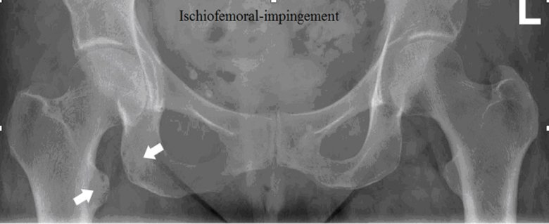
Ischiofemoral impingement

What is it:
– Narrowing of the space between the lesser trochanter of the femur and ischium of the pelvis

Presentation:
– Pain often reported in the lower buttock region, groin/inner thigh +/- snapping/clunking sensation
– Close involvement of quadratus femoris muscle, proximal hamstring tendon and psoas tendon
– Combination of hip extension/adduction/ER can be provocative with closing in of the lesser trochanter of the femur towards the ischium OR stretching of quadratus femoris (opposite of closing down pattern)

image.png

– Long-stride walking can be provocative (again in an ext/add/ER position – lagging leg)
– Clinically subjective reports are load dependent
– Can mimic presentations similar to Lx referral, “sciatica”, proximal hamstrings, groin etc
Imaging:
– MRI findings may show quadratus femoris oedema, narrowing of the ischiofemoral space with coxa valga (although this may also represent standing posture at the time of imaging, may  not be an accurate finding more so just incidental)
– Noted bilat ischiofemoral narrowing doesn’t seem to correlate with bilat symptoms
image.png
 
 
Risk factors:

– Risk factors may be gender (more females reported ?greater Q-angle), following surgery (e.g. THR, femoral osteotomies), or other hip joint pathologies (OA, cam lesions, previous DDH or Perthes), functional patterns (reduced hip abd moments), tendon pathologies (hamstring enthesopathies, psoas etc)

Management:
– Medical Mx

– Surgical resection (esp if cause is tumour, osteochondroma)
– CSI to quadratus femoris if indicated
– Edu re: avoiding impingement positions +/- taping
– NSAIDs
– Ensure any concomitant pathologies addressed
– Correction of any potential LLD if significant (as this may further reduce ischiofemoral space)
– Strengthening of abductors
– Stretching e.g. ERs
– Graduated RTS approach