Author: Michael Ranger

Solving the shoulder

Shoulder

Solving the shoulder.

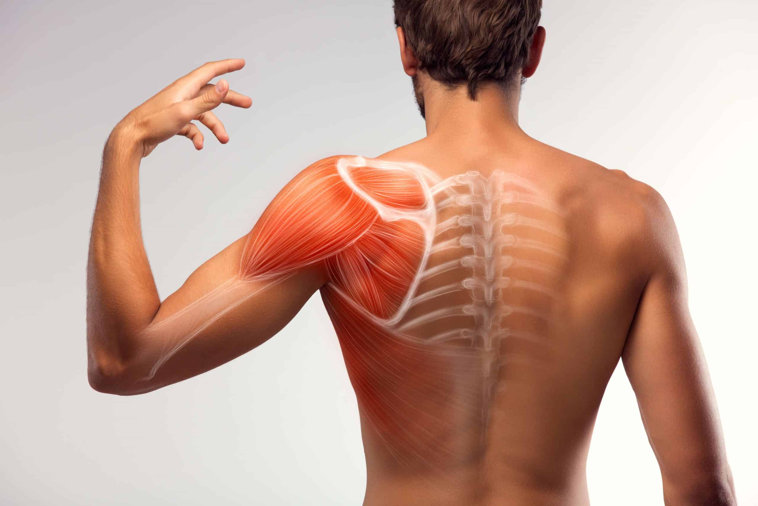
Shoulder pain and injuries are very common. Your shoulder is the most mobile joint in your
body. The reason for this abundance of movement is due to the tiny shoulder contact zone, with
the glenoid fossa (shoulder socket) covering only a quarter of the humeral head (ball). This
makes the joint quite unstable which is why your shoulder muscles are so important to a
normally functioning shoulder to help stabilise the shoulder joint.

Common conditions of the shoulder include:
– Rotator cuff tendinopathy or tears
– Bursitis
– Shoulder dislocation (instability)
– Adhesive capsulitis (frozen shoulder)
– Shoulder arthritis
– Fractures

Rotator cuff pathology
– The rotator cuff is a small group of four muscles that move and control your shoulder
joint. Individuals with a rotator cuff injury will often have a painful arc, pain with overhead
activities, lifting, reaching behind the back, sleeping on the affected side and pain at
night.

Shoulder bursitis
– Shoulder bursitis is due to an inflamed shoulder bursa. The bursa is a fluid-filled sac that
is used as a cushion between tendon-bone and tendon-tendon to reduce the friction and
irritation between them as they move. There are several bursae in your shoulders and
inflammation of this bursa typically results in pain on the outside of your shoulder, pain
with overhead lifting or reaching activities, increased pain at night and pain may spread
down your arm towards the elbow or wrist.

GH instability
– Refers to the inability to maintain the ball of the upper arm bone in its normal position in
the shoulder socket. A dislocated shoulder is where the ball of the upper arm bone is
forced fully out of position which is usually associated with extreme pain and the inability
to move your arm until it is placed back in the socket. Shoulder subluxation is a partial
shoulder dislocation when the shoulder joint comes out partially before relocating itself.
Symptoms of shoulder instability may include repeated instances of the shoulder giving
out, a persistent sensation of the shoulder feeling loose, or “slipping in and out” of the
joint, or apprehension to put your shoulder in certain positions.

Adhesive capsulitis (frozen shoulder)
– Frozen shoulder is a gradual onset of pain and stiffness due to shoulder capsule
inflammation and fibrotic adhesions that limit your shoulder movement. Frozen shoulder
will usually have an insidious onset with a progressive increase in pain initially and later
a gradual decrease in active and passive glenohumeral joint range of motion. It is more
prevalent in women and commonly occurs in individuals 40-60 years of age. Individuals
will often have difficulty grooming, performing overhead activities, sleeping, and
particularly fastening items behind the back.

Arthritis
– Shoulder arthritis is a common source of shoulder pain in older adults. Everyday wear
and tear damages your shoulder joint and the chances of arthritis are more likely if you
have injured or overused your shoulder joint. Exercise is a great way to reduce the
incidence of shoulder arthritis as well as help control arthritic pain, strengthen muscles
and improve your joint and muscle flexibility.

Fractures
– A fractured shoulder is most commonly a fractured humerus which is your upper arm
bone between your shoulder and elbow. The location of these fractures can impact your
treatment because of the bone attachment of your essential shoulder muscles. Your
physiotherapist will be able to explain what you should and shouldn’t do if you have a
fracture in a particular region where there is a muscular attachment.

If you would like more information about different shoulder injuries or you are currently
struggling with shoulder pain, feel free to make a booking today
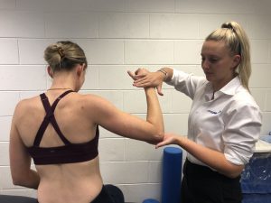
Shoulder rotation

Eliza with patient assessing shoulder range of movement


Written by Eliza Osborn who works from the Physio Plus Footscray clinic.

Staff Profile – Brayden Gaviglio

Brayden is a Mackay local who moved to Townsville to complete his studies at James Cook University. Brayden has a specific interest in orthopaedics, sports and musculoskeletal conditions.
After completing his studies in 2018, Brayden returned to his home town to start his career at Physio Plus. He has since been lucky enough to be able to increase his skills and work closely with a knowledgeable and supportive team.
Outside of work Brayden is actively involved in senior Rugby league as both a player and physiotherapist. Brayden has experience playing and working with both local and representative sides.
Clinical experience in both private practice and inpatient hospital care allows Brayden the knowledge base to give all patients the best possible care whatever their presenting complaint.
Brayden works out of our Willetts centre suites in Mackay and is hoping to combine his studies and local knowledge to provide quality health care to the region.

Adolescent sports injuries

Young athlete

Adolescent sports injuries

There are two types of sports injuries: traumatic and overuse—or acute and chronic. An injury that occurs suddenly, such as a sprained ankle caused by an awkward landing, is known as an acute injury. Up to 60 per cent of all sporting injuries in adolescents affect the ankle.

Chronic injuries are caused by repeated overuse of muscle groups or joints and include cramps, tears and strains. With increased exposure to sport in the adolescence period, there is higher risk of overusing the muscles, tendons and bones.

In adolescent children with a joint injury (sprain), the growth plate of the joint may be injured. The growth plate is the area where the bone grows throughout childhood until the skeleton is mature.

Commonly, the anterior cruciate ligament (ACL) inside the knee can be injured in cutting, landing and twisting sport activity. In contact and dodging sports, there is a reported increase in the amount of these injuries, particularly in girls (up to eight times that in boys).

Return to sport/exercise is varied guided by the area and type of injury sustained. Through consultation with your chosen practitioner, you should plan your return to sport considering all variables to reduce the risk of recurrent or secondary injuries.


What are important measures to take to prevent injury or re-injury?

Before starting a sport, adequate warm up with emphasis on cardiovascular fitness, muscular strength and coordinated skills is important, as well as nutrition and hydration. It is important to avoid heavy fatigue as tired muscles stiffen and lose the ability to freely work, which increases risk of strain during quick activity.

Adequate recovery is also very important. Some adolescent children play for several teams and several sports, increasing their exposure risk to injury. During growing years, it is recommended a child receives a minimum of eight hours sleep and good nutrition to prevent chronic and overuse injuries.

Good equipment such as correct shoe type and fit, breathable sports clothing, mouth guards and helmets are vital in injury prevention. Suitable supervision and umpiring to maintain fair play is also essential for safe play in organised sport and recreational activities.


Young Athlete additional information

For a more detailed look into the intricacies of care of the young athlete, check out the following fantastic link to a resource provided by the AIS: https://www.ais.gov.au/position_statements/content/sport-specialisation-in-young-athletes .


Staff profile – Eliza Osborn

Eliza Osborn

The Physio Plus Footscray team is proud to announce the addition of Eliza Osborn to the Physiotherapy team.

Originally from Gippsland, Eliza graduated from La Trobe University with a Bachelor of Applied Science and Master of Physiotherapy Practice.

Eliza’s passion for physiotherapy stems from her sporting background and keen interest in health and fitness. Personally, Eliza has played and continues to play competitive netball and professionally she has worked with the Melbourne University Blacks in the Victorian Amateur Football Association and has recently gained a position as a Physiotherapist with Williamstown FC in the VFL, assisting with injury prevention and management.

Eliza has experience with a range of musculoskeletal and sport-related injuries. Rehabilitation to optimise function and recovery in all cases is a focus of Eliza’s to help her patients achieve their desired goals in the fastest possible time. Eliza prefers a hands-on approach utilising the best available evidence and manual therapy techniques with a strong focus on exercise rehabilitation and injury prevention.

Outside of the clinic, Eliza enjoys the outdoors – all things boating and water sports, playing basketball & netball and spending time with family and friends.


ACL Injuries

Anterior Cruciate Ligament (ACL) injuries:
what is the usual history?

A good history alone will often diagnose to near certainty an ACL rupture.  This is what you might expect to hear from a patient with acute ACL rupture:

  • Non-contact event usually when decelerating, stopping suddenly, twisting, cutting, or jumping.
  • “Knee falls in” with a fixed foot, creating the sheering which exceeds the capacity of the ACL.
  • Oftentimes the patient will recall hearing or feeling a “pop”.
  • Sometimes they may report brief a hyperextension of the knee joint.
  • There is usually immediate severe pain, which eases over a period of minutes.
  • Patients may then feel they can continue activity, but notice “giving way”.
  • Usually considerable swelling occurs within few hours.
  • In a subacute presentation patients involved in sports may describe feeling like they need to “round” their cuts rather than being able to pivot easily on the injured leg.
  • In a contact injury (typically football) the ACL is usually ruptured after a direct blow to the lateral side of the knee.  Other structures are frequently injured in addition to the ACL. This is often seen as the ‘unhappy triad’ that includes the ACL, medial collateral ligament (MCL), and the medial meniscus.


Anterior Cruciate Ligament (ACL) injuries:
To reconstruct or not?

If there is one injury that strikes fear into the hearts of most active people it is the dreaded Anterior Cruciate Ligament (ACL) rupture of the knee.  It’s likely you’ll be asked, “Should I get my knee done?”

After an ACL injury, it is common for a patient to feel angry, depressed, frustrated and uncertain about their future. Making a plan with your patient is important and provides them with hope. Your plan should include (and in this order):

1. Gathering the rehab team
2. Starting rehabilitation straight away
3. Deciding whether to have surgery (or not)

The rehab team is likely to include exercise professionals (e.g. physiotherapist), a surgeon, a GP/sports doctor, a coach, team-mates, family, and friends.

Patients should begin high-quality rehabilitation immediately after an ACL injury irrespective of surgery plans.

Deciding whether to have surgery is complex.  Encourage patients to gather information, consider their goals, consider their values, and consider risks.  The best course of action is to try to return normal knee function as soon as possible while deciding whether to have surgery.

There is no clear evidence that surgery is superior to undertaking high quality rehabilitation alone. There is only one published randomised trial comparing the two options. This reported no difference in pain, function or return to pre-injury activity levels at 1-, 2- and 5-years after an ACL injury.

It is clear that some patients need surgery and that some will cope well without.  Of course in either case they need physiotherapy and indeed we typically see the surgical group for a longer period.


ACL Rehabilitation: What to expect.

We rehabilitate ACL injuries in phases.  There are many protocols, but three main phases.:

  • Early:
    • Exercises to regain full range of movement and muscle activation.
    • Riding an exercise bike.
    • Practising weight bearing and walking evenly on both sides.
  • Middle:
    • Exercises to single leg balance and control.
    • Exercises to improve general leg strength (eg. squats, lunges, and deadlifts).
    • Exercises to improve single leg strength with (eg. single leg squats).
    • Practising running.
  • Late:
    • Exercises to improve landing ability (eg. hopping and landing practice).
    • Exercises to improve your ability to change direction.
    • Practising unexpected change of direction and agility.
    • Gradually returning to sport-related activities.

There exists a significant risk of re-injury.  We manage this risk by only clearing people to return to sport after they pass a battery of performance, confidence and functional tests and return to sport criteria.  Even after return to sport, we teach exercise based injury reduction programs as part of ongoing risk mitigation.

Of course we are happy to see your patients with knee injuries and to be part of their rehab team.  Referrals can be made by phone, walk-in, online or email found here.

Bowen Summer 10s Carnival

The Bowen Summer 10s Carnival of Rugby took place on the weekend with the Tropic Thunder taking out the Women’s 10s and the Miners taking out the Men’s competition.

Although wet and wild, our very own Senior Exercise Physiologist Yvette Porter competed for the Ladies Carltons playing some technical footy, scoring a few flashy tries and completing a drop kick conversion along the way.

Great work Yvette and everyone that enjoyed the weekend at the Bowen Summer 10s.

Back Pain

Lower back pain – According to the WHO, 60-70% of individuals will experience debilitating lower back pain at least once in their lifetime. This makes it one of the most common presentations we see at Physio Plus.

Given the large prevalence in the community a lot of research has gone into lower back pain. The STOPS trial (specific treatment of problems of the spine) looks at diagnosis and management of lower back pain. The most common sub groups of back pain are outlined briefly below and in more detail via the specific area links.

  • Disc herniation with associated radiculopathy (this is explained in greater detail on our radicular leg pain page).
  • Reducible discogenic/ Non-reducible discogenic lower back pain
    • Common findings we see here are:
      • limited sitting time before pain
      • pain worse the day after the incident
      • difficulty with forward bending, lifting and sneezing
      • history of working in a manual labour job
    • Facet joint dysfunction
      • Common findings of this pathology are:
        • One sided symptoms that tend to be quite localized.
        • Pain with leaning back or side bending to the affected side
      • Multifactorial/non-specific lower back pain (this is explained in depth below in the acute and persistent sections).

If you would like more information about different back injuries or you are struggling with back pain currently, feel free to make a booking today.


Ehlers-Danlos Syndrome (EDS)

What is Ehlers-Danlos Syndrome?

Ehlers-Danlos Syndromes are a group of disorders that share a number of common features such as; easy bruising, joint hypermobility, skin that stretches easily and weakness of tissues.

Ehlers- Danlos syndromes are inherited in the genes that are passed from parent to child. They are categorised according to both the forms of genetic transmission and the varied features.

What are the types of Ehlers-Danlos Syndromes?

The Classical type of Ehlers-Danlos usually presents as joint hypermobility and skin laxity and fragility. The fragile nature of the skin is prone to bruising and tearing easily. Joint instability can lead to commonly occurring strains, sprains and dislocations. This classical type is directly passed on from parent to child.

Hypermobility type is as the name suggests, the type where the major factor is joint hypermobility. Any joint in the person’s body can be affected and results in frequent dislocations.

The vascular type of Ehlers-Danlos syndrome is arguably the most severe as it can cause spontaneous rupture of arteries and bowel which can lead to death. Veins can be visible through the skin and the degree of skin laxity can be varied. Clubfoot can be present at birth. It is most commonly directly passed on from one parent to child, but it can also be recessive meaning that it might only be found in one generation of members of the same family, meaning that the individual must inherit 2 copies of the mutation, one from each parent.

Fragile eyes, significant skin and joint laxity and severe curving of the spine are all traits of the Kyphoscoliosis type. Like the vascular type of Ehlers-Danlos, the kyphoscoliosis type can be passed from parent to child and can be recessive.

Patients with the arthrochalisa type are short in height and suffer from severe joint laxity and dislocations. The level at which the skin is affected by the syndrome is quite varied, and a skin biopsy can be used for diagnosis. Dominant and recessive inheritance occurs with this type.

A rare form of Ehlers-Danlos Syndrome is the dermatosparaxis type. This type can also be diagnosed with a skin biopsy. Patients with this type of syndrome have severely fragile skin which is very soft, resulting in sagging and folding.

Tenascin-X type of syndrome is inherited as a recessive direct passing from parent to child. Patients with this type present with hyper elastic skin, fragile tissue and joint hypermobility.

What are the Signs and Symptoms of Ehlers-Danlos syndrome?

The symptoms include loose skin that stretches easily and skin that is saggy and soft in appearance that may be fragile or tear easily. Bruising easily without a significant trauma to the bruised area and joints that move easily past the usual range of motion are signs indicative of Ehlers-Danlos.

What is the treatment for Ehlers-Danlos?

The treatment for Ehlers-Danlos varies patient to patient depending on the symptoms the individual presents with. Skin protection is critical as the fragile skin can be hard to stitch should injury occur. It is recommended to protect the fragile skin from sun and trauma where possible. As joint injury is common, bracing can be used to aid in joint stability and exercises strengthening the muscles around the joint can help to minimize joint injury.