Author: Michael Ranger

Popliteal Artery Entrapment Syndrome (PAES)

Popliteal Artery Entrapment Syndrome (PAES)

Popliteal Artery Entrapment Syndrome (PAES) is an obscure cause of exercise induced calf pain.
Posterior calf pain is a common presentation in the young athletic population, and so it is important
to recognize that PAES is an uncommon presentation. Therefore, it is important to consult your
regular Physio if you are experiencing ongoing pain, they can perform a thorough assessment to
correctly diagnose your issue. However, our interest today lies in PAES and how its presentation is
very similar to chronic exertional compartment syndrome (compartment syndrome), commonly
referred to as shin splints or calf cramps. This article looks to detail the specific differences between
the two conditions and explore potential treatment options that can be explored.

PAES typically affects a young 20-40yo athletic population and is up to 15x more common in males
than females. PAES is where the popliteal artery becomes dynamically compressed by the medial
gastrocnemius (gastroc) head through exercise. Causes are either anatomical variance in the medial
gastroc insertion or excessive hypertrophy of the muscle. The popliteal artery dilates as the blood
pressure increases in the vessel causing a dynamic compression against the medial gastroc head.
Patients with PAES will typically present with intermittent claudication (posterior calf pain/cramping)
and/or paraesthesia during exercise or exertion. This presentation is almost identical to the typical
compartment syndrome where the calf muscles swell from increased blood perfusion and become
compressed against the fascia. The main subjective difference is that PAES is more specifically
exacerbated by exercise intensity rather than volume. Whereas compartment syndrome requires a
certain duration of exercise to present, PAES can present almost instantly under the right
circumstances i.e running uphill, repetitive jumping, sprinting. Another difference is that athletes
with compartment syndrome will have decreased tolerance to exacerbating factors as a game
progresses whereas PAES symptoms remain consistent relative to intensity.

Differentiation between the two conditions can be determined therefore with a few clinical tests
and confirmed on imaging. Exercises such as repeated hopping on one leg or running can be used to
induce symptoms in the clinic. PAES will present sooner than compartment syndrome with a loss of
the dorsalis pedis or posterior tibial pulse, symptoms also settle a lot quicker as they settle with the
recovery of the athlete’s heart rate and blood pressure. If PAES is suspected, diagnosis can be
confirmed with a dynamic MRAngiogram, where the patient performs their provocative activity and
then the scan is performed. A positive test will show decreased blood flow through the popliteal
artery as it intersects the proximal medial gastroc head.

Early detection and intervention is important as prolonged stenosis of the vessel can cause scarring
and atherosclerosis of the inner vessel wall. If left untreated PAES may require invasive procedures
to stent the vessel open and can significantly impact an athlete’s career. Therefore, early detection is
key. Standard treatment of PAES involves relocating the medial gastroc head, optimizing the
popliteal artery tract. However, botulinum A toxin (botox) injections used locally to the site of
constriction in the medial gastroc head has gained traction in recent literature and is showing
promising results. The procedure is far less invasive than traditional measures and has been shown
to effectively reduce the muscles constriction of the vessel during exercise. Another benefit is that
recovery time is significantly reduced, the player can return to sports almost immediately as function
and pain allow.

Physio management is involved in retraining the muscle to promote muscle atrophy in the medial
gastroc head and guide successful return to sport. Long term management involves educating the
patient to avoid excessive training of calves that would promote increased medial calf bulk which
would encourage the condition to return.

Take home, is that this condition is quite RARE. Your exercise induced calf pain is far more likely to
be a result of the more common diagnoses, such as compartment syndrome. However, the purpose
of this piece is to explore this interesting albeit rare condition and raise awareness of potential
differential diagnoses that exist with exercise induced calf pain. If you are concerned about pain that
you experience whilst exercise, it is best to discuss this with your local physio. They are experts in
diagnosing your pain and providing you with a plan for management and recovery.

Muscle strains, torn muscles, pulled muscles, DOMS?!?! Part 2. Wilson Tang

Muscle strains, torn muscles, pulled muscles, DOMS?!?!

Part 2.

After reading Part 1 you should be feeling a bit wiser on what muscle strains/DOMS can feel like and why they happen. In Part 2 we’ll cover some basics of what to do and how to best kick-start your rehab journey.

Early stages of physiotherapy rehabilitation for muscle strains and DOMS.

Often we find ourselves stuck with questions such as “How long will this take to heal?” or “When can I go back to playing competitive sports?” or “Should I ice it or put a heat pack on it?” Firstly let’s state that there is no miracle cure, no magic wands or special pills that replace healing time and hard work. But that doesn’t mean you can’t make things as comfortable as possible in the meantime. And it also doesn’t mean that you can’t do other things to keep you physically active and happy.

DOMS recovery

When looking through research literature, the jury is still out when it comes to what works best in minimising DOMS/recovery time. Interventions can include cryotherapy (ice or cold exposure), massage, compression, stretching, massage guns, creams, and a whole plethora of other knick knacks you can think of. Be cautious with anything that promises or sells miracure cures (remember there aren’t any). The evidence that does exist suggests that most of the aforementioned interventions may provide some pain relieving effects short term, but anything past that is still not 100%.

For many individuals with DOMS, management may simply mean getting back to some light mobility work or gentle exercise, or even just exercising another part of your body. This is one reason why many individuals like to isolate body parts (think leg day, arm day etc) during gym sessions, that way they allow rest days during the DOMS recovery period.

To hot pack or cold pack?

I find heat packs and ice packs are hugely personal preference (some people love heat packs, some people love cold packs). If we are getting technical and looking at the physiological mechanisms then usually a heat pack for muscle relaxation and to increase blood flow, and ice packs to help hinder swelling and inflammation. A recent study by Jerrold Petrofsky and his colleagues in 2015 looked at heat and cold therapy both immediately after exercise and 24 hours after exercise, and its effects on strength, pain levels and several other measures. Their results indicated that both were useful for different things at different times.

Imaging or no imaging?

Imaging is not required for DOMS, however if soreness does persist for more than several days even with resting then a quick visit to your physiotherapist may be warranted. With strains, mild strains don’t often require imaging unless symptoms are persistent. Given imaging can be expensive and findings of imaging may not actually change rehabilitation management, many healthcare professions would recommend against imaging for low grade strains early on. Moderate strains may require imaging again depending on the recovery process so far, as well as the site of strain.

Severe strains typically do require imaging, as this will help clarify whether further referral to an orthopaedic specialist is necessary.

It should be noted that imaging is a great tool to rule out any nasty pathology, but doesn’t tell you where pain is nor how capable you are. Use imaging sparingly.

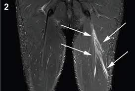
The above picture shows an MRI of a low grade hamstring muscle strain, where the muscle fibres show some oedema (swelling) but fibres are predominantly intact.

Courtesy of Aspetar Sports Medicine Journal (https://www.aspetar.com/journal/viewarticle.aspx?id=28#.YmnEDNpByUk)

Strains

For mild muscle strains the recovery process may take several days to several weeks, but you may be able to continue light or gentle exercise. It’s often a good idea to ease off intense sport or the specific exercise which caused the muscle strain to begin with, as you don’t want to keep hammering that same injured muscle whilst it’s healing. Maintain mobility and movement, but keep effort low, simply easing back the intensity/effort can do the trick with mild strains.

Moderate muscle strains will take longer, we are talking several weeks to potentially months. As mentioned before this will be influenced by the location of the injury. For these injuries there may be a period of relative rest, and usually you will have to “baby” the injured area for a good while before going back to your exercise. Mild analgesics (such as paracetamol) or anti-inflammatories may be required to help with symptoms. If there is some muscle tension or tightness after the healing process is complete, gentle stretching may be introduced but avoid this during the early days of recovery.

Severe muscle strains will take the longest, and recovery can be months if that. In these cases imaging is usually advised to help determine if surgical input is required.

Anti-inflammatory medication or not?

There is discussion amongst healthcare professionals nowadays regarding the use of anti-inflammatory medication during early stages of soft tissue injuries. It is argued that disrupting the inflammatory process during early stages of a soft tissue injury may interrupt the body’s natural physiological reaction to an injury. What happens is during the inflammatory stages (usually lasting for several days after an injury) the body floods the injury site with cells that limit further injury and kick-starts the healing process. Thus stopping this would appear counterproductive to healing and optimal tissue repair.

HOWEVER, given inflammatory pain can be quite significant and limit the amount of active recovery/rehab you may partake in there should always be a balance between using medication for symptom relief when needed, and avoiding it if possible.

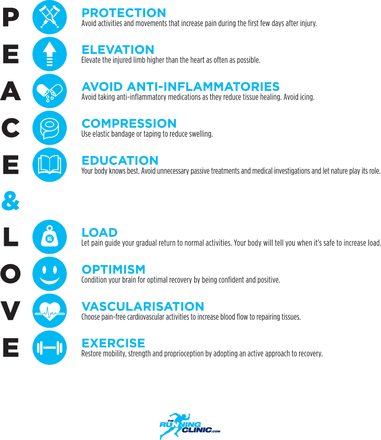
Courtesy of Dubois & Esculier (doi.org/10.1136/bjsports-2019-101253)

TIPS FOR DOMS AND MILD TO MODERATE MUSCLE STRAINS.

Exercise:

  • Early loading and movement is key! Talk to your physiotherapist and find movements that your body likes or can tolerate, and avoid the ones that aren’t so great for the time being. This may include targeting all your muscles apart from the affected one (but may also include targeting the affected one in a reduced manner).

  • We know your body heals best and feels best when you move, gone are the days of absolute bed rest!

  • Mobilise early and avoid stretching until later stages of the healing process if necessary.

Pain relief:

  • Medications can be a great way to make the healing process a little more comfortable (think paracetamol and ibuprofen, always consult your GP or pharmacist for medication information).

  • Heat/cold therapy.

  • Creams/sprays – Topical creams can provide temporary pain relief (think brain distraction), with some topical creams also having anti-inflammatory properties as well.

Diet:

  • Keeping a balanced diet that is nutrient rich and contains adequate protein will help fuel the body for recovery.

Protection:

  • In some cases taping, bracing or splints may be used during early stages of an injury, but remember you want your muscles and body to do the work in the long run and not the tape/braces/splints. Chat to your physiotherapist about best taping/bracing/splint options for your specific injury.

Later stages of physiotherapy rehabilitation

A common mistake with muscle strain rehabilitation is stopping when pain has fully resolved, after all pain is a great motivator! However, by the time muscles have healed and you are pain-free it is likely the muscle has weakened or deconditioned to a degree. Often we prematurely return to a high level of activity without doing the necessary work to get our injured muscle back to its former strength, if not further. As you can imagine this alone is a risk factor for a repeat muscle strain, and so the vicious cycle starts. Take your rehab a few steps further to really set yourself up brilliantly for the long run.

Making sense of all this and putting all the rehab principles into practice can definitely be a daunting task. To get your rehab on track for the best outcomes possible, book in to see your local friendly physiotherapist.

Part 2 SUMMARY

  • For DOMS, don’t worry! Keep going with gentle exercise, avoid high intensity stuff involving the sore muscles.

  • After being cleared for a severe strain, GET MOVING! Start low and slow and work within low/no pain movements.

  • Medications can help make the process more comfortable, but healing takes time. Be patient.

  • Keeping active will best help the healing process, and help retain muscle flexibility/range, cardiovascular endurance and muscle function.

  • Fuel your body with adequate protein and a variety of nutrients/food groups.

  • Imaging only when necessary.

  • Once you think you’re out of the woods, take it several steps further for re-injury prevention.

Muscle strains, torn muscles, pulled muscles, DOMS?!?!  – Part1. Wilson Tang.

Muscle strains, torn muscles, pulled muscles, DOMS?!?!


Part 1.

We’ve all heard of the common old “I’ve strained/pulled/torn a muscle” or even the basic “Ohhh the muscles are a tad sore after that workout!”. But what has actually happened when you get a strained/torn/pulled muscle? And why do we experience DOMS (delayed onset muscle soreness) after a good workout? Knowing what happens during a muscle strain is the first step in understanding how to best manage your muscle soreness or injury, and will get you set up for the best outcomes both short- and long-term.


Basic muscle anatomy

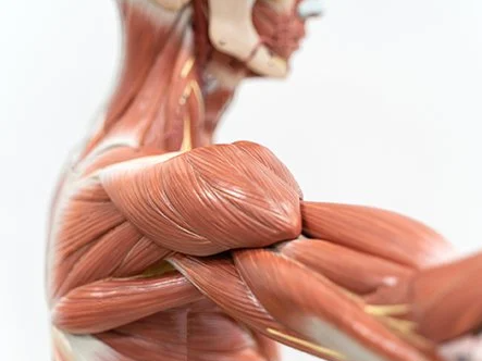
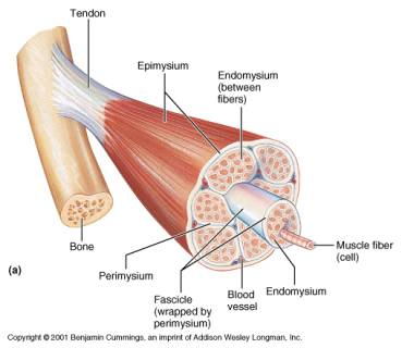
To understand muscle strains and DOMS we must first appreciate that muscles are complex and adaptable things. Picture muscles as a bunch of tubes (called sarcomeres) bundled together like many straws, which attach from bone to bone via tendons (see Image 1). Muscles (red part) have a stretchy, elastic-like property (think strong elastic band) and do all the hard work. Tendons (white part) on the other hand don’t do as much work, but act more to transfer force that the muscle produces, and connect muscles to bone. Tendons are strong, flexible, and play an important role as well.


Image 1. Cross section of a muscle


How does a muscle work?

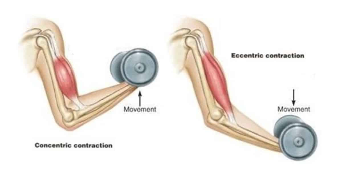
Muscles are living, breathing (sort of) things which work by pulling (concentric contraction) and releasing (eccentric contraction). Picture a bicep curl. Your bicep works concentrically to lift the weight up and bend at the elbow, and works eccentrically when slowly lowering the weight back to a straight arm position (see Image 2). In technical terms muscles work via the ‘sliding filament theory’ involving actin and myosin units which form the sarcomeres. Both concentric and eccentric muscle contraction requires varying degrees of effort, and this is where muscle injuries and soreness starts to come into play.


Image 2. Concentric and eccentric contraction of the biceps muscle.


Why do muscle strains occur???

Muscle strain injuries ultimately result from tissue/muscle failure. In other words, the muscle is exposed to a load or force higher than they can handle, and the activity demand exceeds the muscle capacity.


Exposure to load/force can come from a variety of sources. This includes lifting heavy objects, to repetitive movements (for example running), to high velocity moments (for example changing direction during footy). Exposure to load/force can lead to small micro-tears within the muscle, and if the force is great enough or prolonged enough then larger sized tears can obviously occur. Remember, since muscles are living/breathing things so they can be slowly trained to adapt to load. This is where DOMS comes in!


What are DOMS?

DOMS refers to that generalised muscle ache we get after a good exercise session, or after attempting a new exercise your body isn’t quite used to yet. I’m sure many of you can remember a time where you have gone back to the gym or a sport after holidays, and the next few days feeling pretty darn sore for the next few days. Then gradually over a few sessions your body gets used to said exercise and that soreness slowly goes away. That is ADAPTATION in a nutshell.


To explain this in slightly more technical terms, during an exercise session you are exposed to high or repeated external forces causing small micro-tears to your muscles, but in a manner that is controlled, not overly painful, and within your muscle tissues capacity (think lifting weights, or doing gentle cardio). Following the cool down period after your exercise session, the body begins the healing process and its associated inflammatory reaction to heal these micro-tears and rebuild your muscle tissue stronger than before (ADAPTATION).It is believed that these micro-tears and the inflammatory response (and several other theories) are what causes DOMS. The peak of DOMS typically sets in 1-2 days after the session is completed, and the soreness begins to ease after this.


It should be worth noting that you do NOT need to experience DOMS in order for adaptation or hypertrophy (building muscle size) to occur. However, the more you exert and challenge a muscle differently the more likely DOMS will occur.


What does a muscle strain/DOMS feel like?

With muscle strains individuals may describe a painful strong pulling sensation, which can appear sharp in nature and progress to more of a strong achy feeling over time. In more significant cases a strong pop may be felt. During early stages there may be limited movement around the injured area, and you may notice some bruising and swelling develop over time. Pain is most likely worsened when attempting to use the affected muscle.

Likewise, DOMS can often result in limited movement but to a lesser degree, and symptoms typically ease with gentle movement. Strains are often felt at the time of exercise, particularly the more severe ones, whereas DOMS is felt over the next few days.


Why do some muscle injuries heal quicker than others?

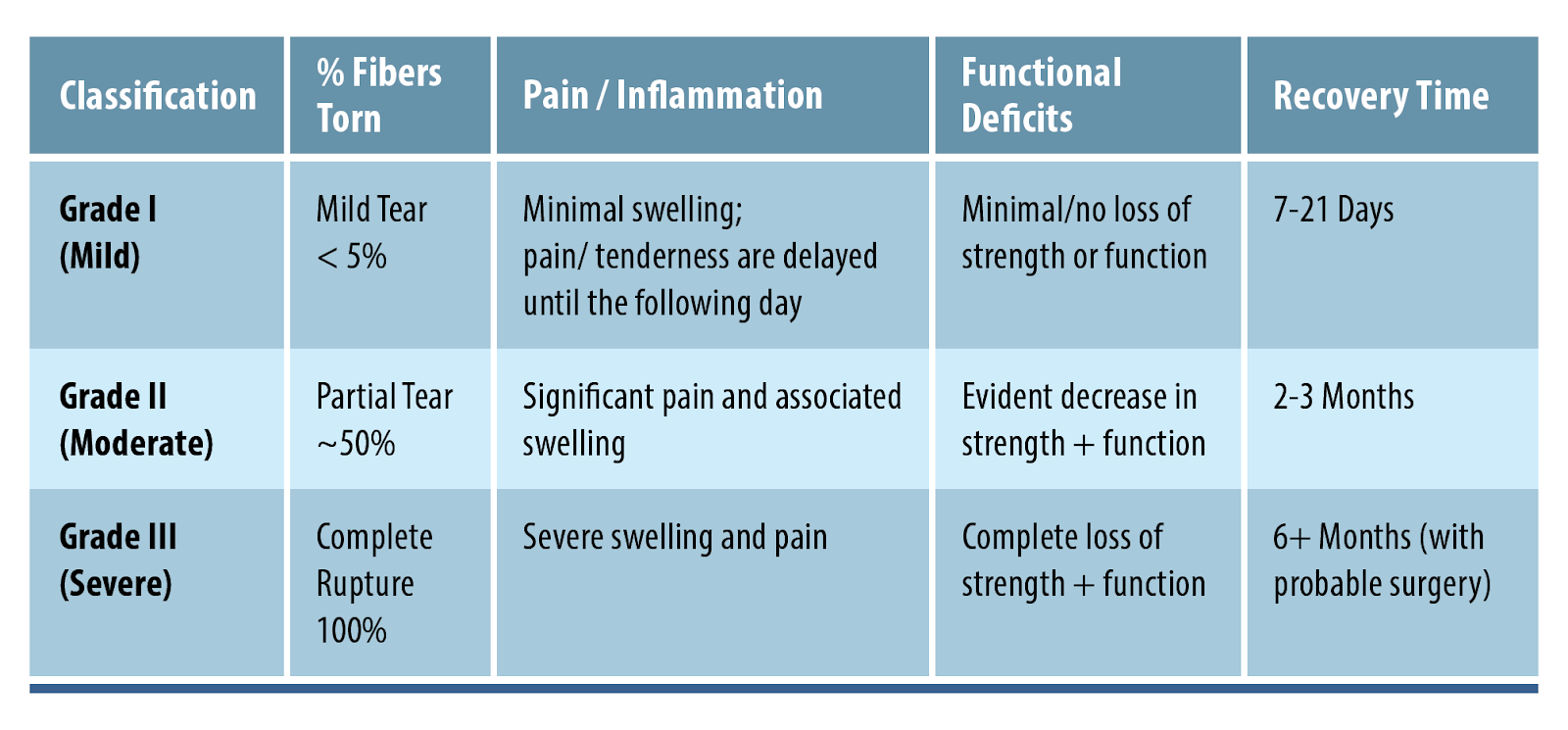
Healing rates of muscle injuries are largely dependent on the location. The injury can occur in three distinct locations; the periphery of the muscle (myofascial), in the bulk of the muscle (musculotendinous), or in the tendon (intratendinous). The most commonly injured location is the musculotendinous part. Naturally, the extent of the injury will also influence how long it takes to heal. Muscle strains can be graded using different scales, although the simplest and most commonly used grading system ranks strains as shown below.


Other comorbidities such as diabetes or smoking can notably slow down healing rates as well.


Part 1 SUMMARY:

  • Sore muscles after a big workout or different workout is normal!

  • DOMS typically occurs the next few days following a workout, whereas a strain is typically felt at the time of exercise.

  • Muscle strains typically occur when the demands of a muscle exceed its capacity.

  • Muscles are adaptable to load, training them up slowly and steadily is your best bet.

  • Healing times vary between degrees of strain, and personal factors as well. Make your own journey!


An early and thorough assessment following a muscle strain is essential whether you are wanting to simply get back on your feet or return to high level sports. Book in to see your physiotherapist for a thorough assessment and tips on how to best handle your individual rehab journey.

___________________________________________________

WILSON TANG, PHYSIOTHERAPIST

Hemiplegic Migraines

Hemiplegic Migraines are a rare type of migraine disorder which involve single sided weakness and aura/sensory symptoms. This can be due to genetics or can occur spontaneously in certain individuals.


Symptoms can range in intensity and can last for a few days and in severe cases a few months. The motor symptoms will tend to last longer than the headache symptoms and these will often present quite similar to a stroke. In the majority of cases patients will tend to make a full recovery.

Triggers can be things such as stress, lack of sleep, emotional distress and physical exertion. Women are three times more likely to be affected and average age of onset of symptoms is between 12 and 17 years of age.

Diagnostic criteria for Hemiplegic migraines is as follows:

At least 2 attacks fulfilling the criteria 2 and 3

Aura consisting of both of the following:

  • Fully reversible motor weakness
  • Fully reversible visual, sensory and/or speech/language symptoms

At least 2 of the following 4 characteristics:

  • At least one aura symptom spreading gradually over at least 5 minutes, and/or at least 2 symptoms occurring in succession
  • Each non-motor symptom lasts 5 to 60 minutes, and motor symptoms last less than 3 days
  • At least one aura symptom is unilateral
  • Aura is accompanied or followed by a headache within 30 minutes.


Given how similar presentations are to a stroke or TIA, CT’s, MRI’s and spinal punctures may be needed to rule out any other pathologies.

The attacks are managed with preventative medications which are taken ongoing for the life of the individual. Though as patients get above the age of 50 the occurrence of motor symptoms tends to reduce and they are more likely to be affected only with migraine and aura symptoms.

Motor symptoms can be treated similar to that of acute stroke rehab and provided all scans are clear of any tissue damage there should be full symptoms resolution in an accelerated time frame.

Stroke rehabilitation

STROKE

A stroke occurs due to disrupted blood supply to the brain. There are two types of strokes that commonly occur ischaemic (blockage of a blood vessel) or haemorrhagic (bleed from a blood vessel). Both will lead to damage to the nerves and cells within the brain. This damage then leads to the functional deficits that are seen in stroke survivors. Physiotherapy has an important role to play to improve these deficits and help the individual get back to living as independently as possible.

Clinical guidelines suggest that where practical patients should commence mobility exercise within 48 hours, therefore in the early stages physiotherapy intervention becomes crucial. A physiotherapist will conduct a thorough assessment of the patient’s level of motor and sensory function and use this assessment to guide rehabilitation requirements. Once rehabilitation is started it is also recommended to complete as much as practically achievable. The more that patients can undertake the better the overall recovery will be.

As part of this rehabilitation process it is recommended that specific goals are set. These goals should be made collaboratively and should involve the client, their family and other treating practitioners apart the health care team. This will help individually tailor rehab programs and keep everyone on the same page with treatment.

For more information check out out full breakdown of stroke and best evidenced care at https://physioplus.com.au/stroke-recovery/ or book an appointment with one of our fantastic clinicians today.


FOOTSCRAY – WE’RE MOVING!

With the redevelopment of the Whitten Oval soon to move into full swing, our suite of over 12 years for Physio Plus Footscray is soon to be flattened.

As such, this has given us the opportunity to move to a new custom fitted out facility just down the road at 1/1 Whitehall Street, Footscray.

We’re looking forward to the new facility being open to you all and being able to welcome you into an open, inviting and clinically impressive space.

Upskill your calf

A calf raise or heel raise is a common exercise prescribed by physiotherapists as part of the rehabilitation in a range of lower limb conditions. At times this can be performed poorly and as such will not have the desired result. Research conducted by Rio et al 2016 focusing on the calf raise as part of Achilles tendinopathy rehab suggests that the simple use of a metronome can lead to a reduction in tendon pain as well as an increase in our control over the muscle (referred to as motor control).


Exercises are normally used as part of rehabilitation to create physiological changes to the muscle with the goal of improving strength. When combined with an external cue (such as a metronome) this begins to target motor control as well. To complete any task successfully you need both the strength and motor control aspect of the movement. This is particularly important in tendinopathy rehab as the pain associated with tendinopathy has been shown to lead to abnormal tendon loading and changes in neural drive (messages from your brain to the muscle both of which affect our motor control. These changes are part of our body’s protective response.


The use of a metronome to pace the concentric (rising up onto your heels) and the eccentric (lower down to the floor) phase has been shown to restore a normal pattern of tendon loading in those individuals with Achilles tendinopathy. This will also lead to an increase in time under tension during your repetitions and thus improvements in the physiological changes within the muscle. As a starting point, set your metronome at 60 beats per minute and aim for a two-beat rise, a two-beat pause at the top and a two beat lower for each repetition. Though this has been predominantly researched in calf raises, there is a further investigation into its applicability for other exercises.


Rio E, Kidgell D, Moseley GL, et al Tendon neuroplastic training: changing the way we think about tendon rehabilitation: a narrative review

British Journal of Sports Medicine 2016;50:209-215.